
”The 3D model allowed us to simulate the surgical procedure, enhancing our perspective, increasing intraoperative safety, and avoiding unnecessary extended resections.
Dr. Emilio PeñaConsultant Colorectal Surgeon, Hospital General Universitario Reina Sofía, Murcia
Clinical Case
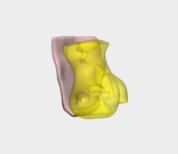
3D Surgical Planning
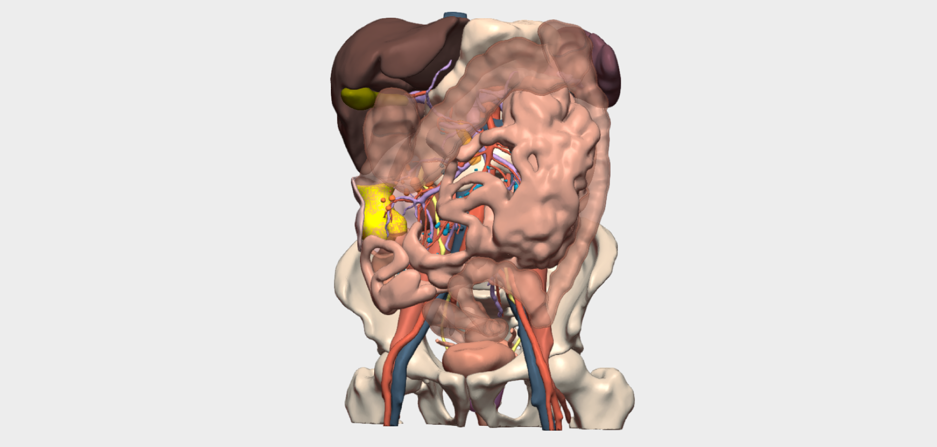
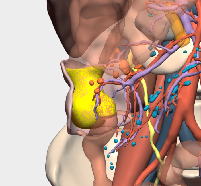
“The 3D model provided crucial information regarding the tumour’s relationship with the retroperitoneal fascia, contrary to what was suggested by CT imaging. This allowed more accurate definition of the retroperitoneal resection plane without the need to include adjacent structures.”
Dr. Emilio Peña
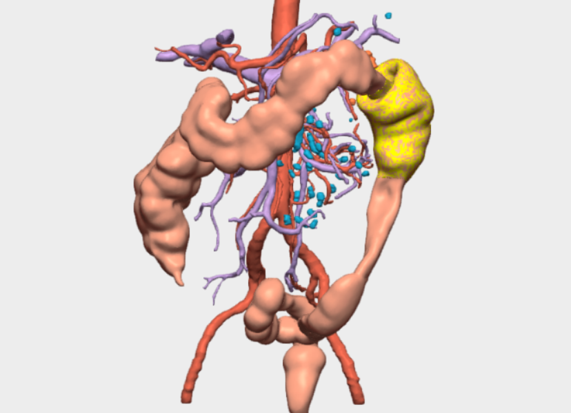
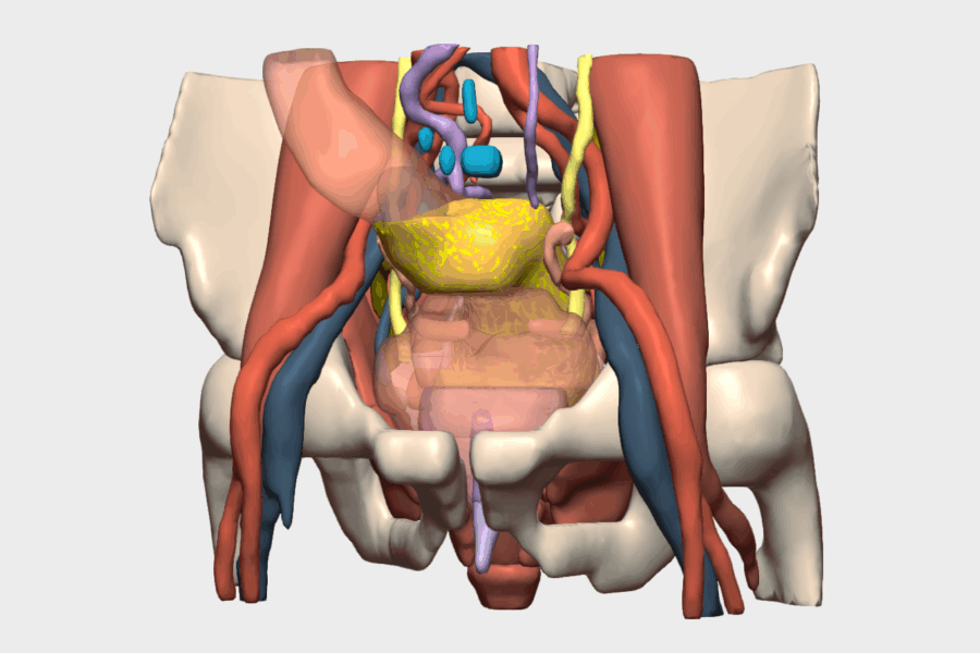
In addition, three-dimensional reconstruction of the venous vasculature revealed a rare anatomical variation, which significantly facilitated the planning and safe execution of the D3 lymphadenectomy.
Value of the “Relationships” Tool
Use of the “Relationships” tool within the 3D model confirmed tumour contact with the retroperitoneal fascia without evidence of infiltration. Tumour involvement of the ileocaecal valve and terminal ileum was also identified, findings that directly influenced the surgical strategy.
Surgical Outcome
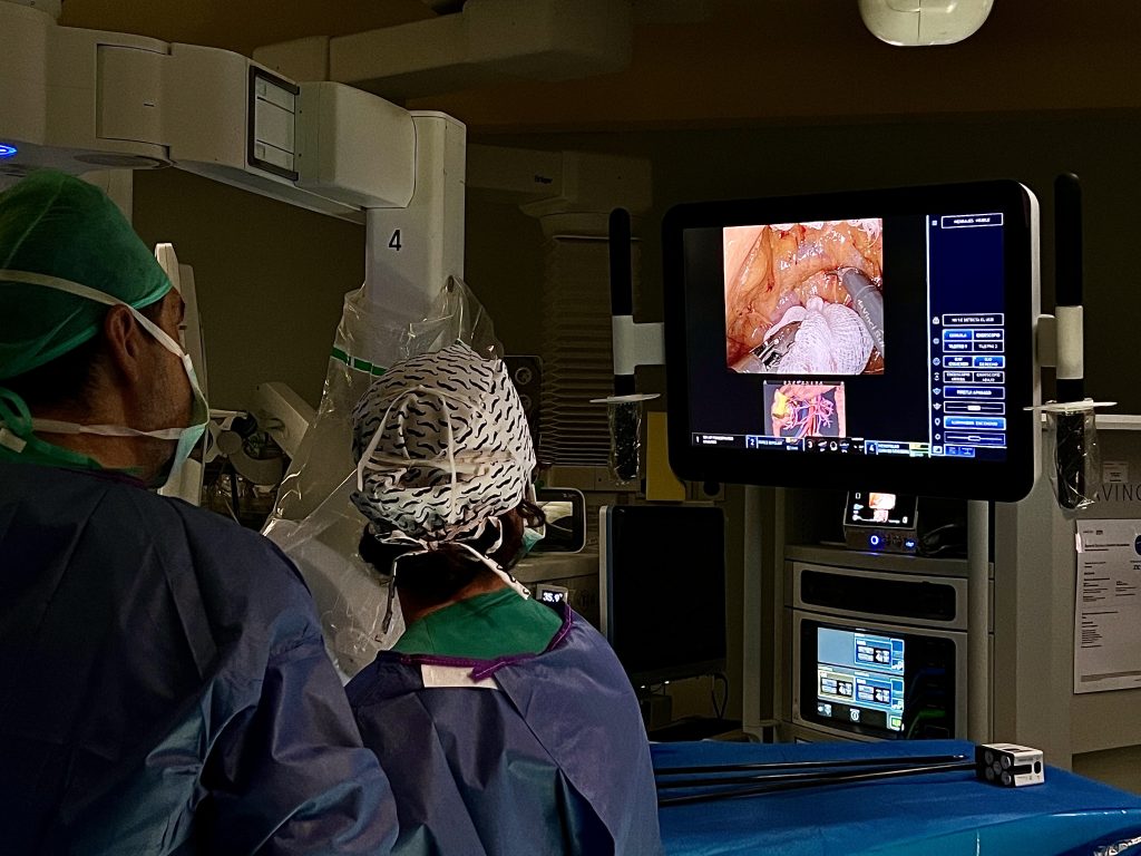
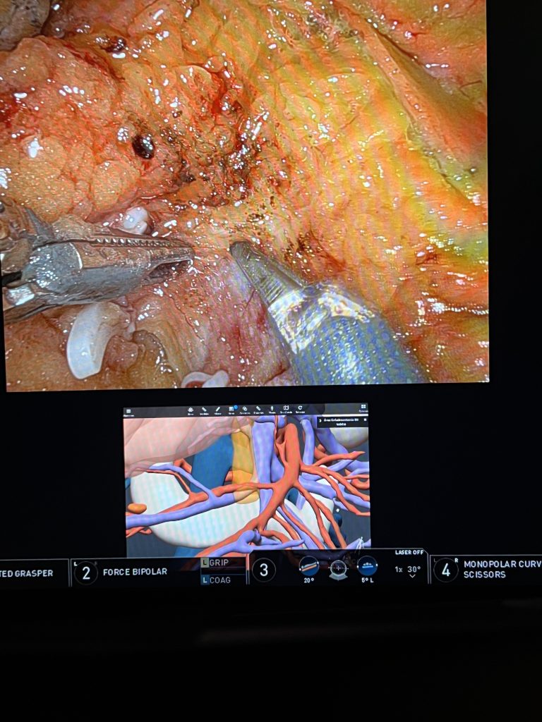
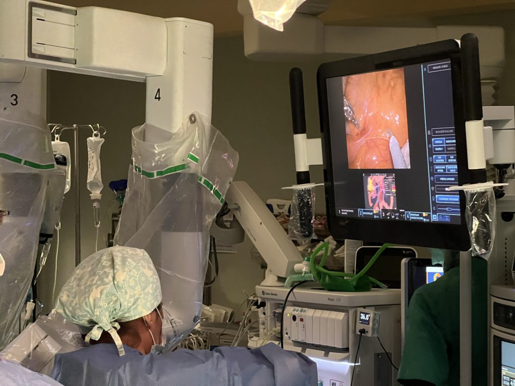
The 3D model proved to be a key tool in preoperative planning, enabling a deeper understanding of the case, enhancing procedural safety and avoiding unnecessary extended resections. Furthermore, it supported a more precise lymphadenectomy by revealing the patient’s vascular anatomical variant.