
Adapted to the hospital’s needs, committed to optimising surgical work and improving outcomes in soft tissue surgery. Most importantly, it increases the quality of care and patient safety.
3D models for surgery: Advanced technology for precise and personalised planning
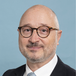
Reconstructions validated by radiologists that optimise surgical planning and increase safety in complex soft tissue procedures.
Endorsed by opinion leaders | +15k surgeries supported
Next -Generation 3D Modelling

The most advanced 3D surgical planner, without software or complications
Our solution enables surgeons to anticipate risks, support clinical decision‑making and minimise intraoperative complications. Designed for complex surgeries, our patient‑specific 3D models provide a complete 360° understanding of anatomy.
Cella Medical Solutions
0:34 duration
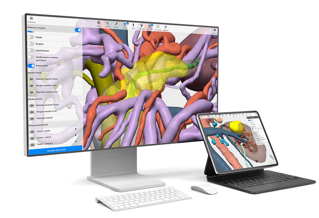
Pre-operative surgical planning
Interact and work on the patient's 3D anatomy
Advanced tools
Automatic calculations
Share models
Save information
Clinical value of 3D models in surgery
- Precise understanding of anatomy
- Anticipate surgical complications
- Reduction of surgical and operative preparation times
- Improved communication and coordination between surgical teams
- Reduce hospital costs and improve efficiency
- Less invasive surgeries with lower patient’s risk
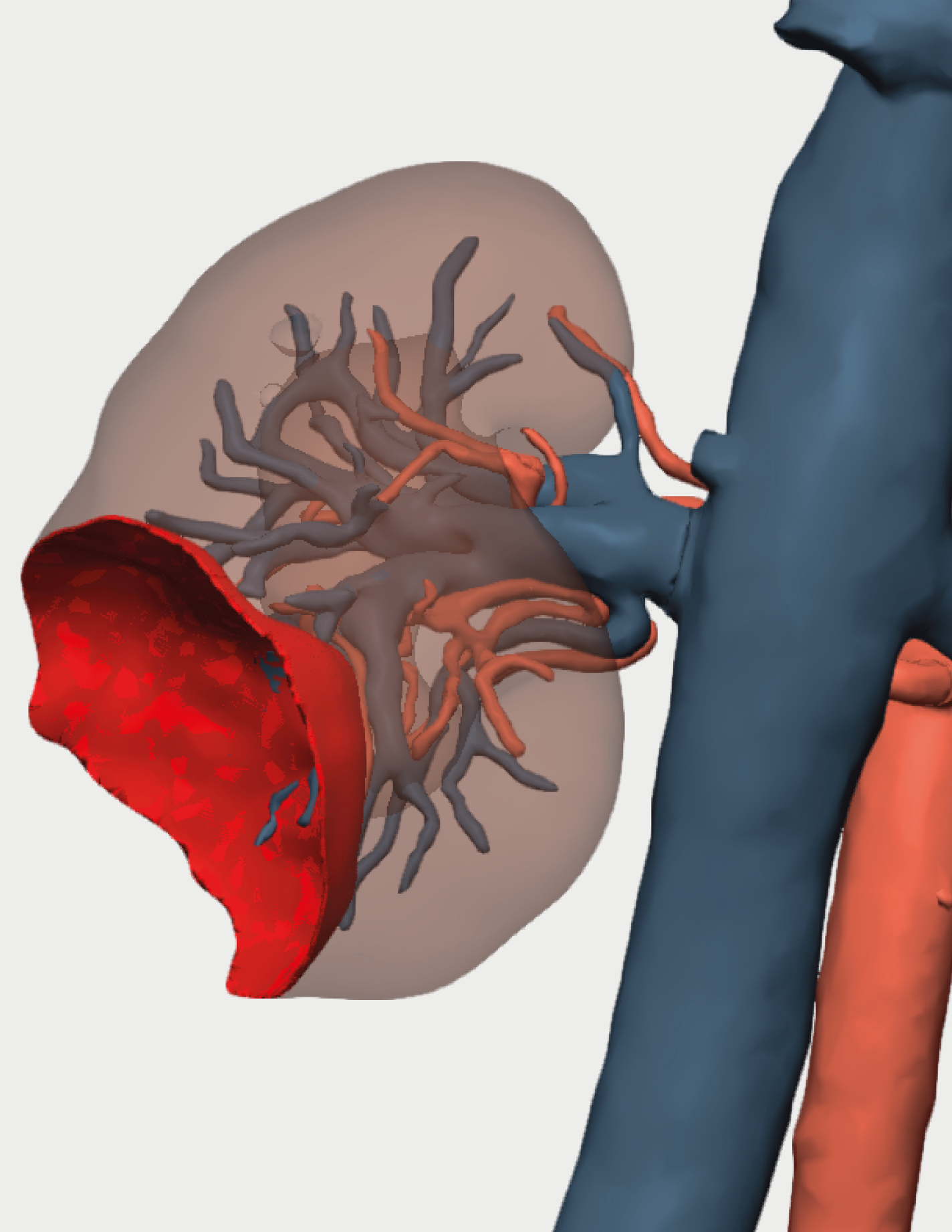
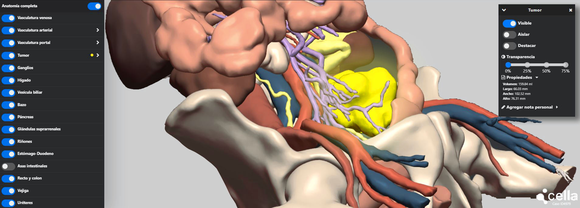
Advanced tools to define the optimal surgical plan for each patient
- Studies of anatomical variants.
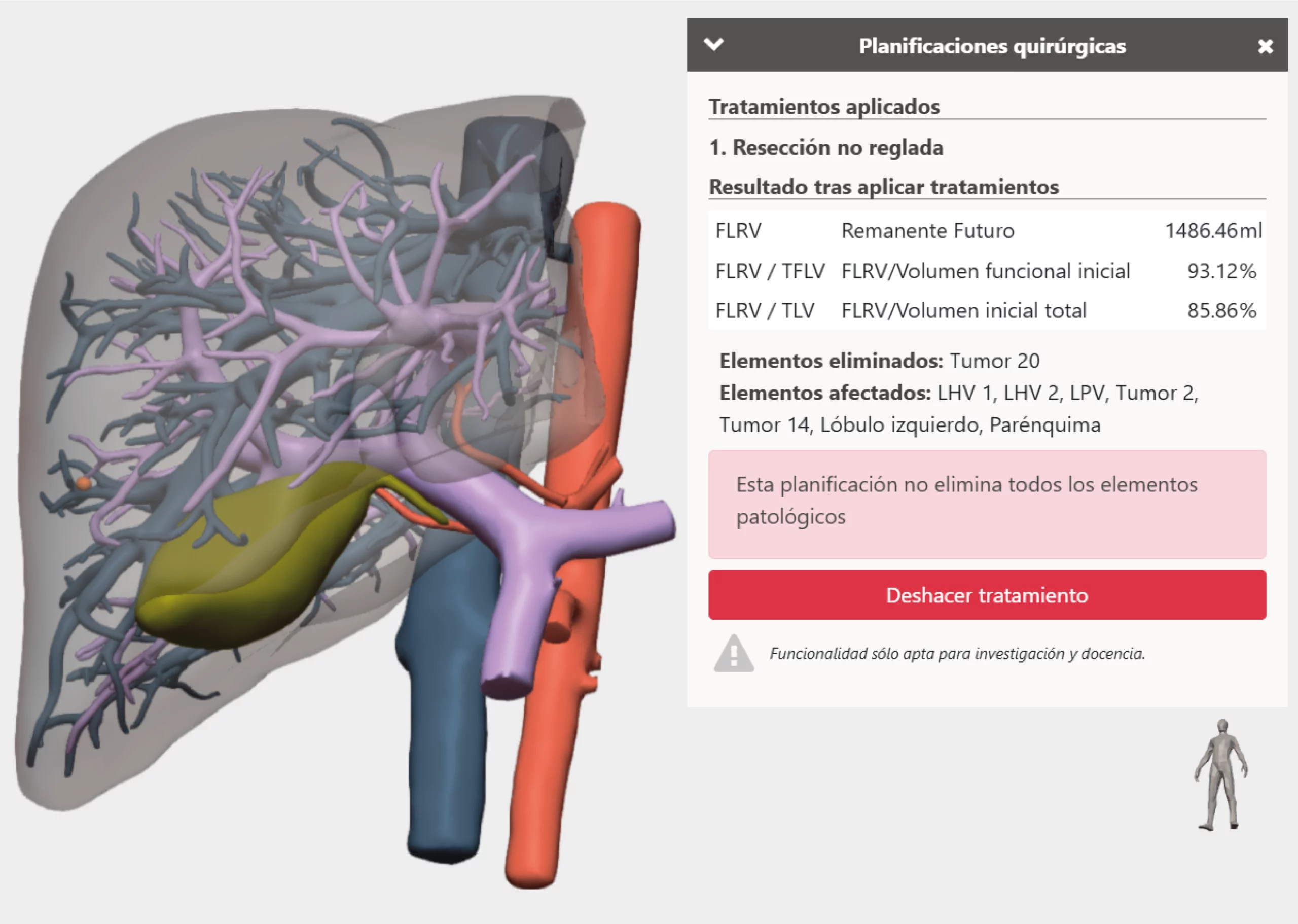
- Planning of regulated and unregulated resections and tumourectomies.
- Simulation of clamping.
- Vascular assessment studies.
- Measurements, calibres and volumes of elements.
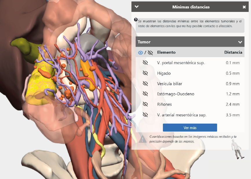
- Calculation of the minimum distance between elements and the lesion.
- Safety margins for tumour resection.
- Virtual navigation through ducts or vasculature.
- Vascular territories.
- Segments: hepatic, pulmonary, renal, etc.
- Infiltrations/contacts between tumours and other elements.
- Surgical bed and exophytic and endophytic elements.
A comprehensive solution for the hospital
Preoperative planning
Specialised support
Intraoperative guidance
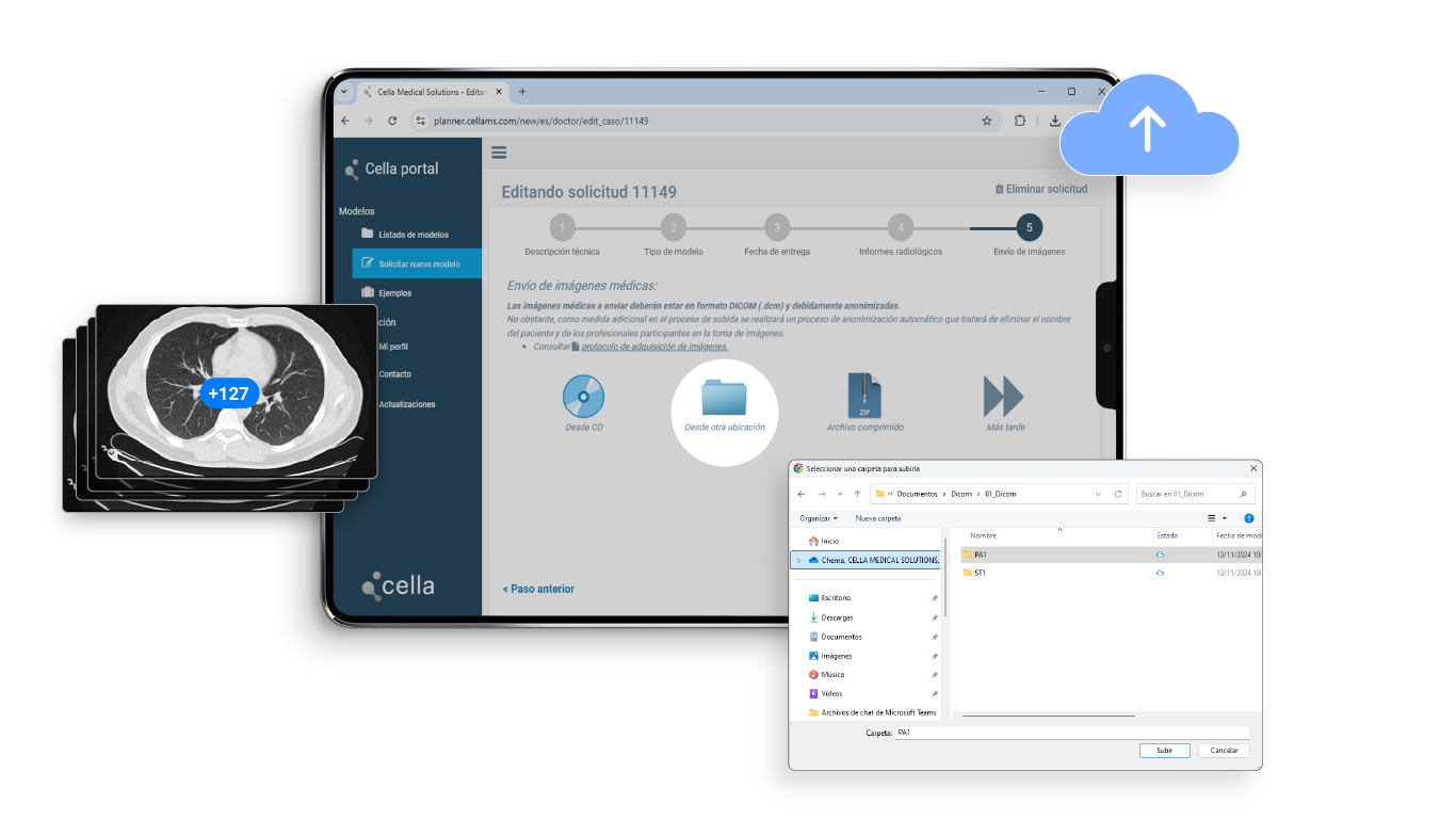
Automated medical image sharing
Preoperative planning
Our web-based planning environment includes advanced 3D modelling tools.
Intraoperative guidance
3D models are seamlessly integrated robotic consoles or laparoscopy towers, providing real-time visual support during surgery.
Automated medical image sharing
Integrating the system with the hospital’s RIS/PACS enables radiological studies to be transferred faster and more easily.
Specialized support
Clinical and technical guidance for hospital teams in the use of 3D models.
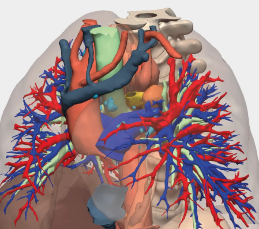
We create 3D anatomical reconstructions from clinical data
What is medical image segmentation?
This is an essential step in 3D modelling, in which anatomical structures such as organs, tissues and blood vessels are identified with precision.
At Cella, we combine advanced image processing technology with the clinical expertise of our technicians and radiologists to achieve highly accurate segmentation.
What makes our 3D models stand out?
We specialise in advanced medical image processing, combining cutting-edge technology with the expertise of our multidisciplinary team, comprising radiologists, engineers, and medical imaging technicians.
Our firm commitment to research and development (R&D) drives us to conduct research in areas such as artificial intelligence (AI) and radiomics, exploring new solutions for surgeons.
Advanced image processing: AI, radiomics and algorithms
Medical image fusion
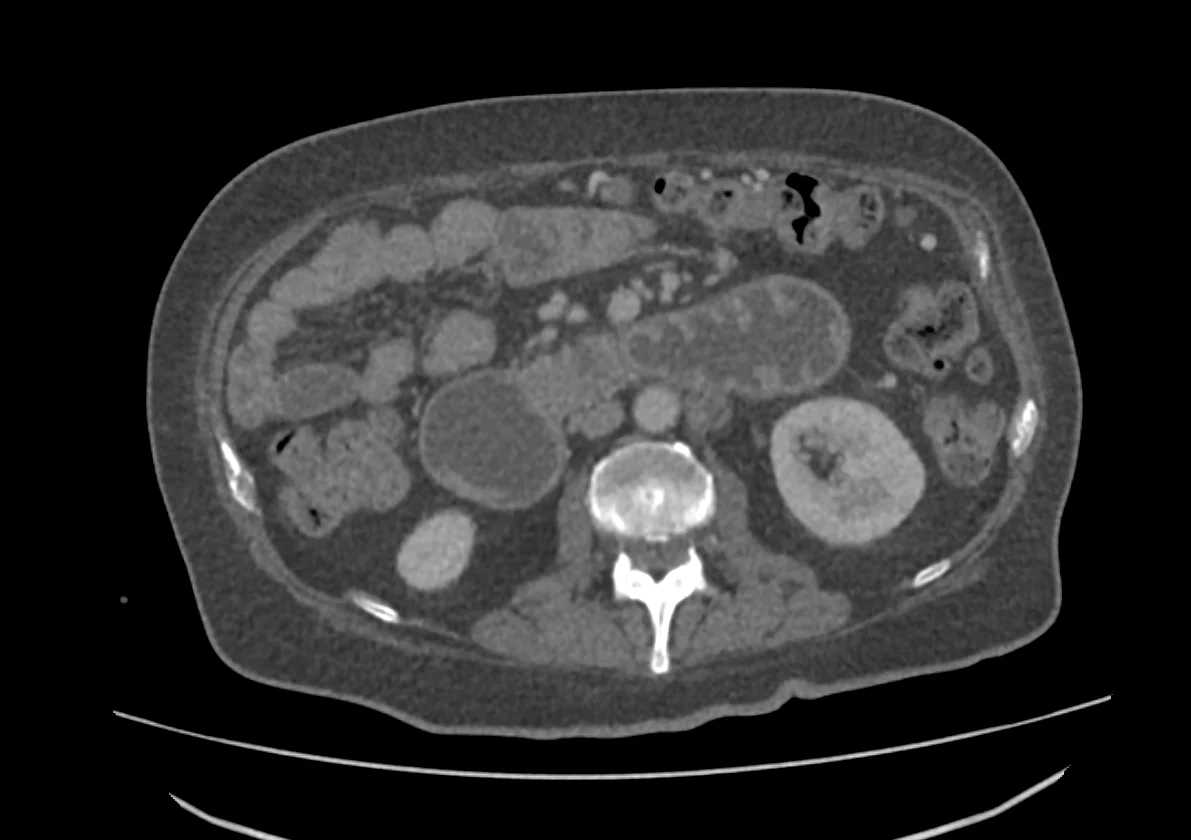
Accepted medical studies: CT, MRI and PET
Clinical validation by radiologists
Surgeons have validated the use of 3D models in clinical practice
+15.000 complex surgeries performed 


Cella complies with the highest regulatory and quality standards
We are committed to establishing and maintaining a Quality Management System that meets or exceeds the requirements of UNE-EN ISO 13485:2018 and the applicable regulatory requirements for medical devices.
Cella Modelling System is a customised 3D model with medical device licensing. Cella Surgical Planner is a visualisation tool for Cella Modelling System.
Frequently Asked Questions (FAQs)
How will I receive the requested 3D models?
To access the requested virtual models, log in to the Cella web platform using your username and password. This will give you access to all your cases.
Is it possible to share virtual 3D models with other professionals on the platform?
Yes. You can create a profile for the entire service or associate a 3D model with multiple doctors, enabling them to work with it from their own profiles.
Are you complying with the Organic Law on Data Protection (LOPD) when sending patient images?
Once the hospital has obtained the patient’s informed consent, the medical images required for creating the 3D model are uploaded to Cella’s web platform. During this process, any identifiable patient data is anonymised and linked to a unique ID to ensure privacy. Cella then assumes responsibility for processing the patient data.
Is it possible to request an urgent 3D reconstruction?
Our emergency management system enables us to process model requests within a timeframe ranging from four days before surgery to 48 hours prior. It is essential for the Cella team to be able to respond to the needs of the centres that rely on our solutions.
Quality excellence
The 3D models adhere to strict quality standards, as evaluated and approved by the UNE-EN ISO 13485:2008 Quality Management System standard for medical devices.
Customer Support
We are here to help with any questions you may have about the application process or our platform. We offer technical support and advice on using 3D models.

© 2026 Cella Medical Solutions