Next Generation
3D modelling
SUCCESS STORY
Dr. Paul Efraín Solís
Hospital General de México
Clinical Case
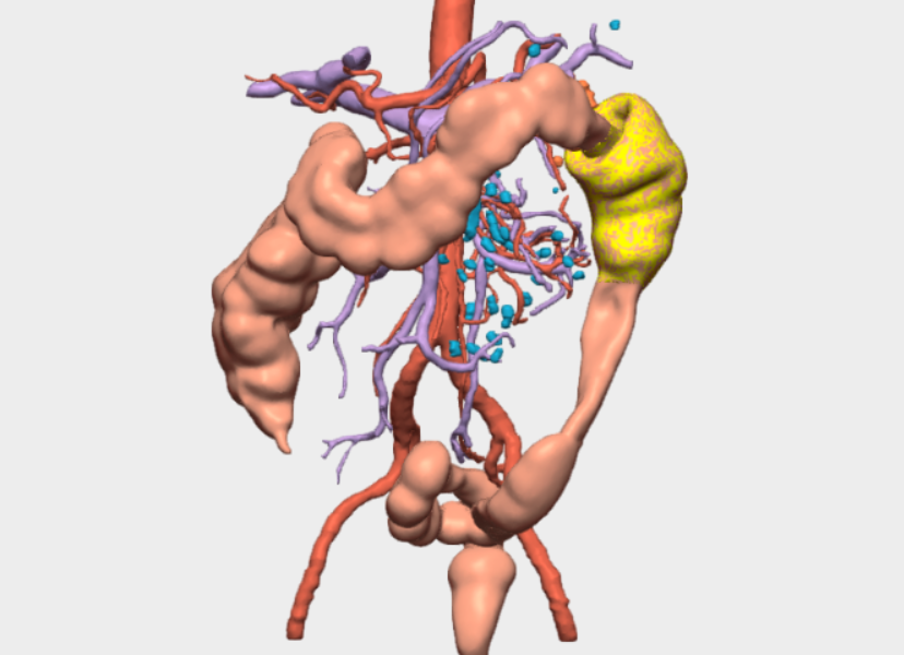
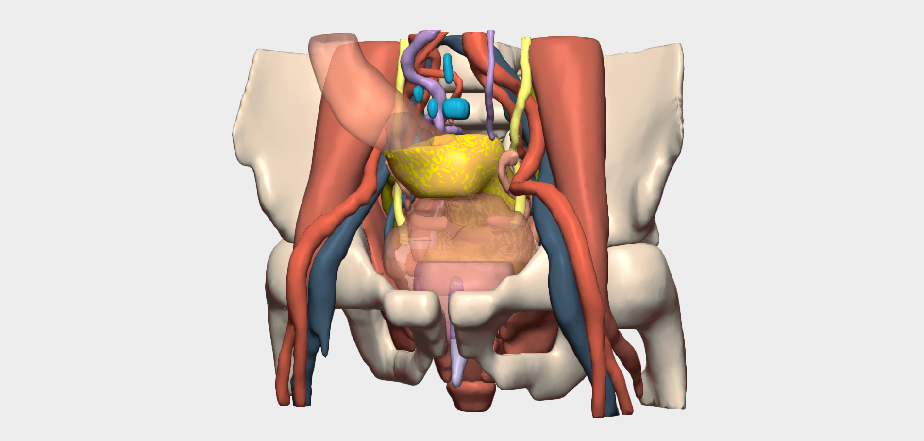
This case involves a 64-year-old male patient diagnosed with a mid-rectal tumour tumour located in close proximity to critical anatomical structures, including the prostate, urinary bladder, and major arterial and venous vessels.
Given the high anatomical complexity, the procedure was planned using a patient-specific 3D model and managed by a multidisciplinary surgical team comprising Dr Paul Efraín Solís Hidalgo, Dr Billy Jiménez, and Dr Juan Antonio Villanueva.
Key Areas of Interest in the 3D Model
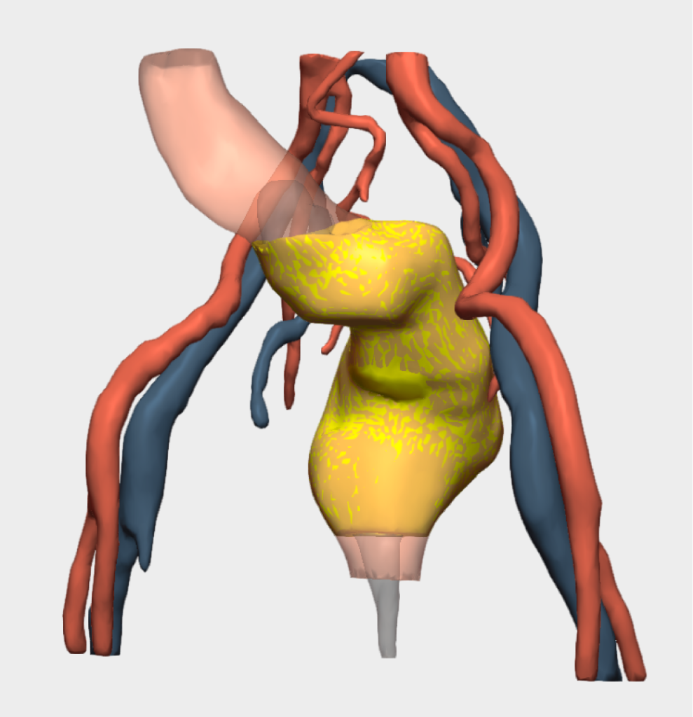
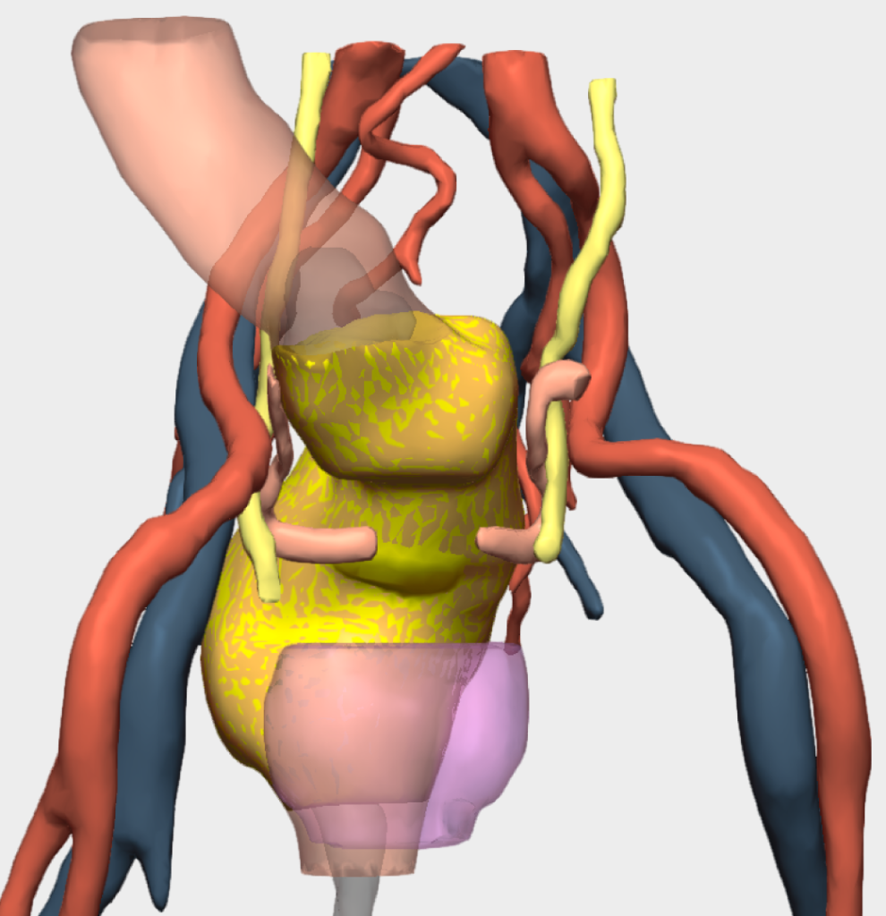
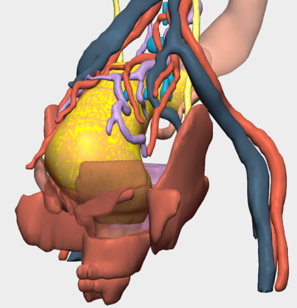
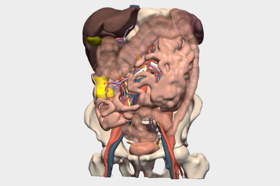
The surgical team highlighted the value of detailed three-dimensional analysis of both arterial and venous vasculature, as well as the pelvic floor musculature. In addition, the 3D reconstruction of the patient’s anatomy proved essential for accurately assessing the tumour’s proximity to the ureters and for clearly defining safe dissection planes, a critical factor for precise and safe surgical planning.
Clinical Value of the 3D Model
According to the hospital team, the 3D model was invaluable in providing a clear and comprehensive understanding of the tumour and its relationship with surrounding structures, enabling accurate evaluation of dissection planes.
The model’s “Planes” tool was particularly useful, allowing visualisation of anatomical sections in the sagittal, coronal and axial planes, similar to conventional medical imaging, but with the added benefit of a true three-dimensional perspective.
Conclusions
The 3D model enabled the surgical team to simulate the procedure in a three-dimensional environment, improving spatial understanding and enhancing intraoperative safety. This approach contributed to optimised clinical outcomes, reduced surgical risk, and the successful resection of the tumour.
”The precision with which the model was developed, and its close resemblance to the patient’s anatomy, is truly impressive. An excellent tool for performing safer surgery.
Dr. Paul Solís HidalgoColorectal Surgeon, Hospital General de México

© 2026 Cella Medical Solutions