
”Given the complexity of the case, the 3D model provided by Cella was extremely helpful in enabling accurate surgical planning
Dr Jennifer Illana WolfConsultant Thoracic Surgeon, Hospital Universitario Puerta del Mar, Cádiz
Sternal chondrosarcoma is a malignant tumour originating from the cartilage of the sternum. It is characterised by its limited response to conventional treatments such as chemotherapy and radiotherapy, making surgical resection the primary therapeutic option.
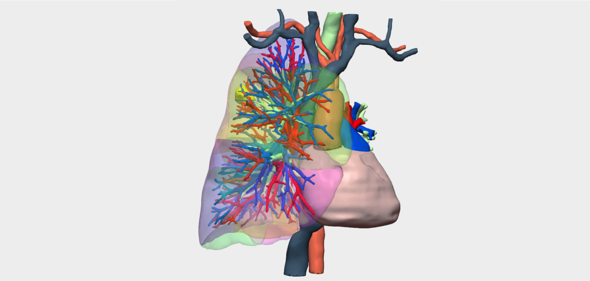
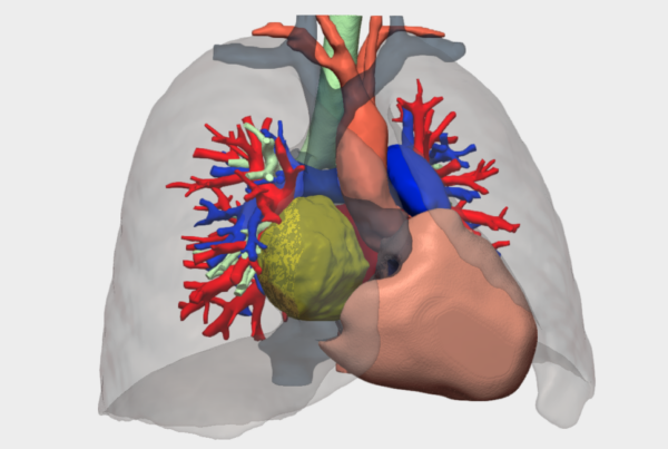
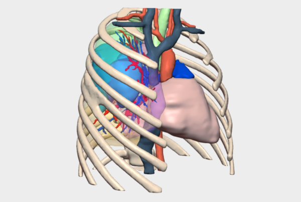
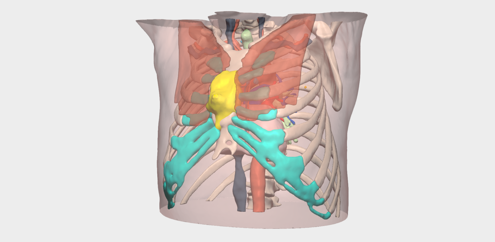
Due to the anatomical complexity of the sternal region, 3D reconstruction–based surgical planning plays a critical role in the management of sternal chondrosarcoma. Patient-specific 3D models enable precise definition of resection margins, improve understanding of the tumour’s relationship with adjacent structures, and support safer surgical decision-making, ultimately contributing to better oncological and functional outcomes for the patient.
Clinical Case Presentation
We present the case of a 69-year-old male patient with a sternal mass that had been present for over one year. Magnetic resonance imaging (MRI) suggested a possible chondrosarcoma. The patient was therefore referred to the Sarcoma Unit at Hospital Virgen del Rocío for further assessment of a suspicious sternal lesion. A core needle biopsy (CNB) was performed, and histopathological analysis initially reported an enchondroma. As a result, no further management was pursued at that centre, and the patient was subsequently referred to the Thoracic Surgery Department at Hospital Universitario Puerta del Mar (Cádiz) for surgical treatment.
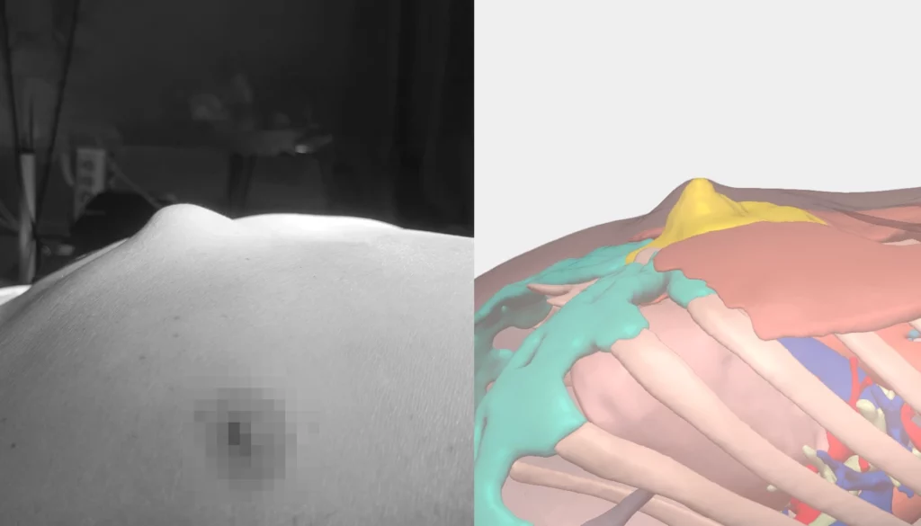
This case posed a significant surgical challenge, as complementary imaging studies revealed that the sternal body lesion had a substantial soft tissue component, with features suggestive of infiltration of the pericardial fat.
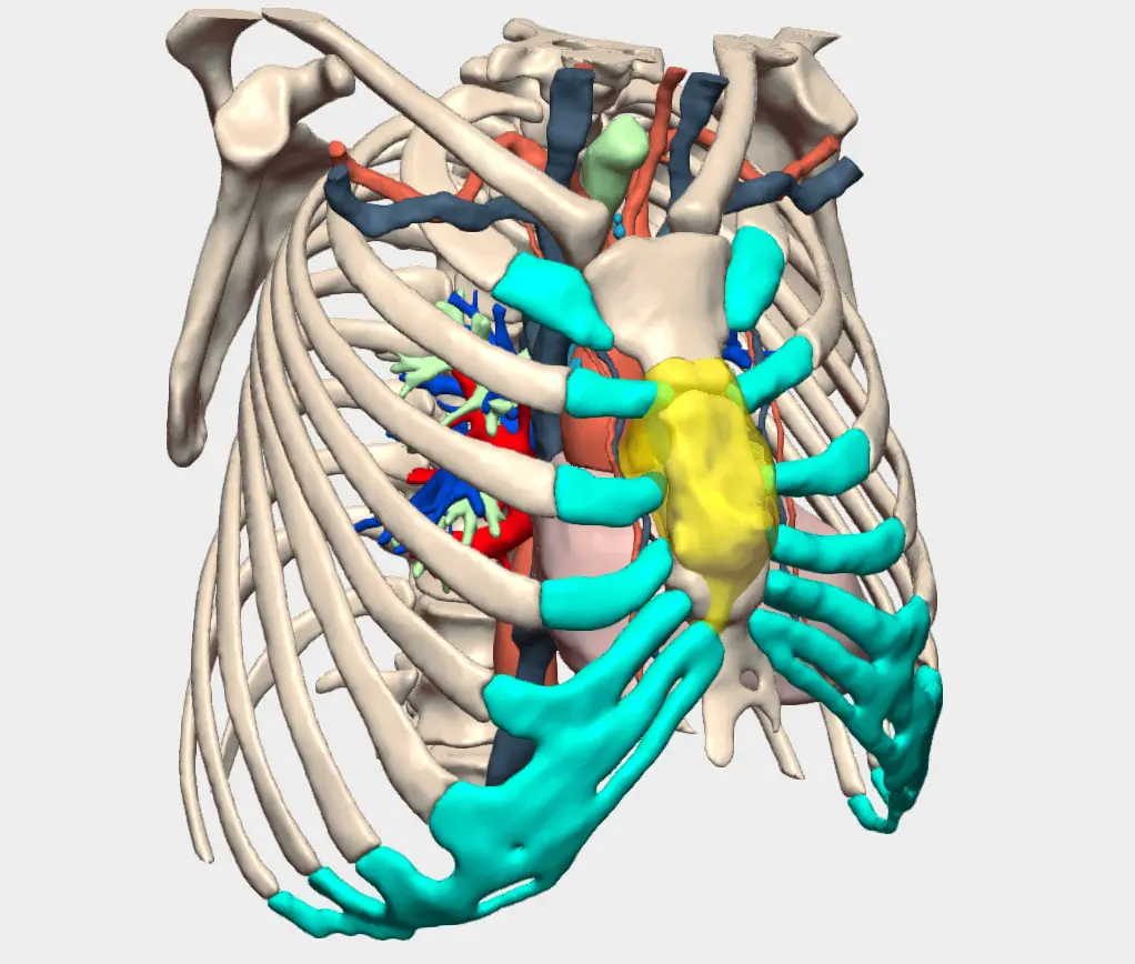
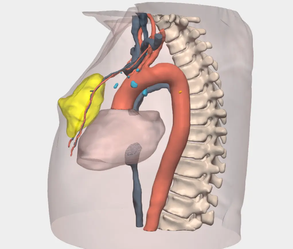
Surgical Planning of Sternal Chondrosarcoma with a 3D Model
Given the complexity of the case, the patient-specific 3D model provided by Cella Medical Solutions proved highly valuable for accurate surgical planning. The model enabled detailed visualisation of the patient’s anatomy and a precise understanding of the tumour’s relationship with surrounding tissues. By using the model’s functionality to selectively add and remove anatomical structures, the surgical team was able to define the optimal resection strategy, supporting complete tumour excision while minimising potential complications.
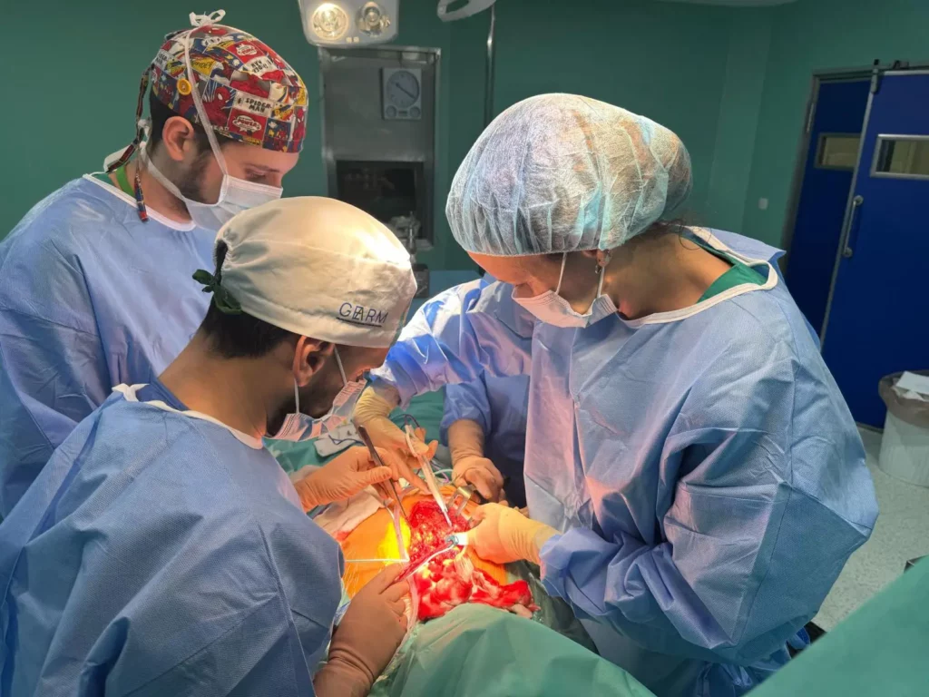
Surgical Outcome and Conclusions
An excellent surgical outcome was achieved. The tumour was completely resected with clear margins, followed by reconstruction of the chest wall defect and sternal osteosynthesis using a sternal plate. Definitive histopathological analysis confirmed the diagnosis of chondrosarcoma, in line with the initial clinical suspicion.

This case highlights the value of Cella’s 3D models as a key support tool in complex thoracic oncological surgery, contributing to improved surgical planning, reduced procedural risk and a more personalised approach based on the patient’s unique anatomical characteristics.