Next Generation
3D modelling
Success Story
Dr. Emilio Vicente
Hospital Universitario HM Sanchinarro
Clinical Case
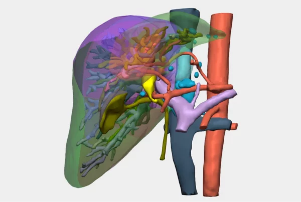
3D Model of Situs Inversus | Cella Medical Solutions
This case involves a 62-year-old male patient with symptomatic cholelithiasis, presenting two highly relevant clinical features that significantly increased surgical complexity:
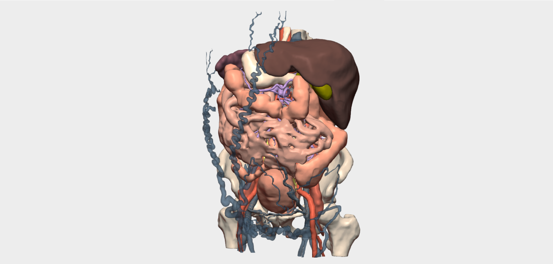
- The patient has situs inversus totalis, a rare congenital condition of genetic origin that affects the spatial arrangement of internal organs, positioning them in a mirror-image configuration. This anatomical variation can complicate surgical procedures, as standard anatomical landmarks are reversed. In this context, accurate knowledge of the exact location and real spatial orientation of the organs becomes essential for safe and effective surgical planning.
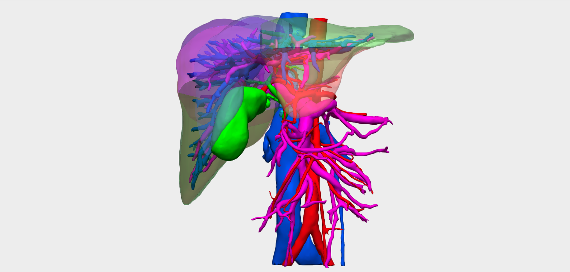
2. The patient also presented with extensive collateral venous circulation within the subcutaneous tissue, associated with a hypercoagulable syndrome and a known inferior vena cava thrombosis. This condition significantly increases the risk of intraoperative bleeding and requires meticulous handling of the patient’s vascular anatomy, making precise preoperative planning even more critical to avoid unexpected complications.
The patient had previously undergone a left transverse laparotomy, as well as a prior procedure involving the inferior vena cava. These factors further complicated surgical planning due to potential anatomical distortion and postoperative changes.
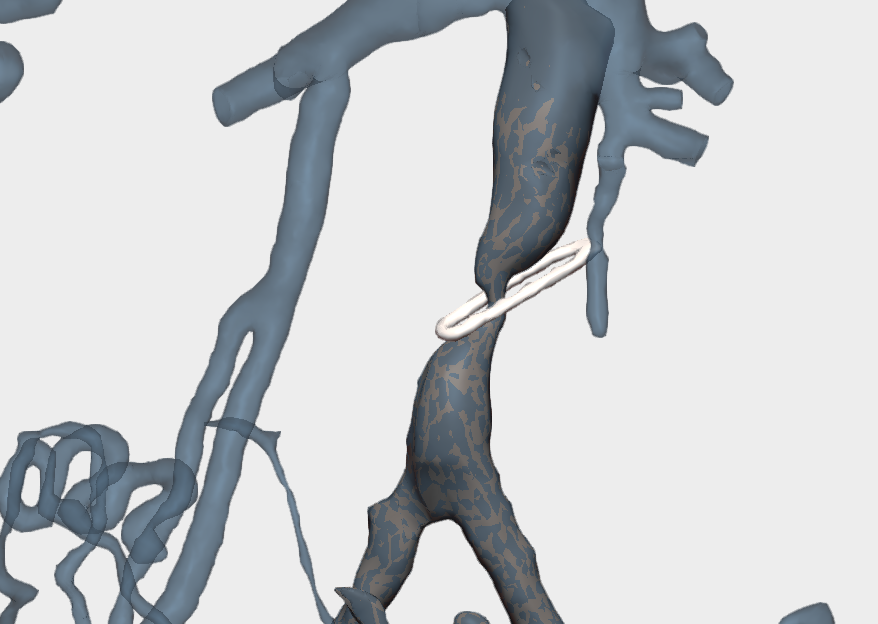
During analysis of the 3D model, the medical team identified an unexpected foreign surgical object within a vascular stenosis. This incidental finding introduced an additional layer of complexity, as it could alter venous haemodynamics and increase procedural risk. Consequently, the surgical strategy had to account not only for the treatment of cholelithiasis and existing conditions, but also for appropriate management of this unexpected finding.
3D Surgical Planning
As highlighted by the clinicians involved in the case, detailed knowledge of the venous vascularisation and its anatomical course was particularly valuable for planning trocar placement.
Three-dimensional planning enabled comprehensive visualisation of the patient’s unique anatomy, allowing detailed analysis of surrounding structures and critical surgical points. The 3D model not only helped anticipate potential challenges, but also improved team preparedness, ensuring that all members of the surgical team shared a clear and accurate understanding of the patient-specific anatomy and the operative strategy prior to entering the operating theatre.
”This model is highly educational and, when planning incisions, it is extremely useful for taking the complex vascular anatomy into account.
Dr. Emilio VicenteDirector of the Department of General and Digestive Surgery, Hospital Universitario HM Sanchinarro