Next Generation
3D modelling
Success Story
Dr. Norberto Cassinello y Dr María Lapeña
Hospital Clínico Universitario de Valencia
Clinical Case
Patient-Specific 3D Model | Cella Medical Solutions
We present the case of a 55-year-old male patient with a medical history of arterial hypertension and left nephrectomy, in whom an incidental finding was identified on computed tomography (CT).
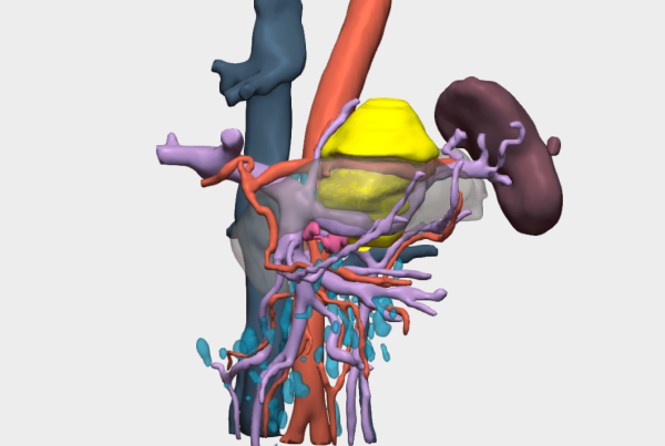
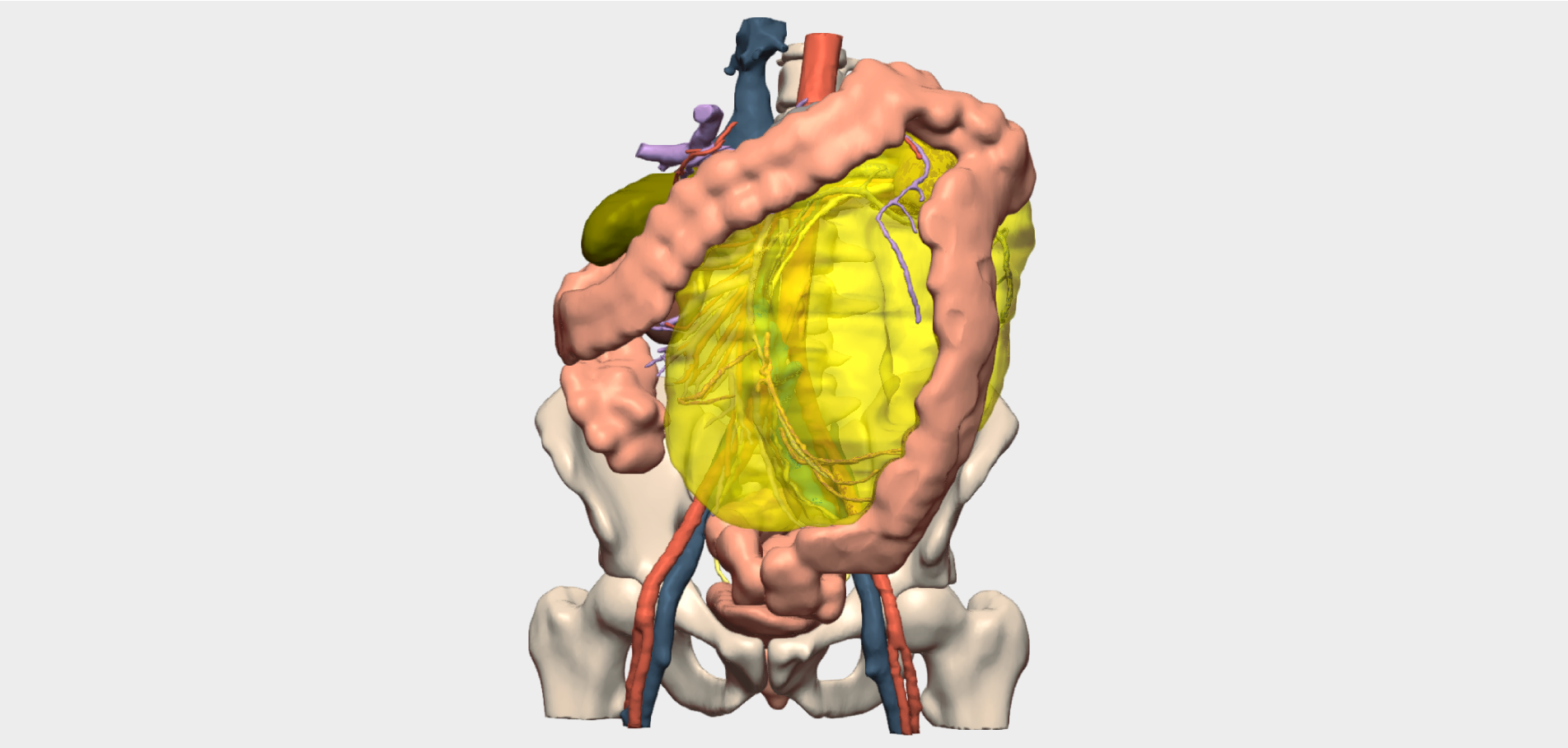
Imaging revealed a solid–cystic retroperitoneal lesion adjacent to the right adrenal gland and in close contact with the inferior vena cava, raising significant concerns regarding surgical complexity and vascular involvement.
Value of the 3D Model in Surgical Planning
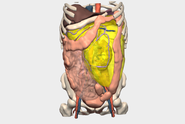
Given the complexity of the case, the Endocrine Surgery team at Hospital Universitario de Valencia requested a patient-specific 3D anatomical model to precisely localise the lesion and clearly define the optimal surgical approach.
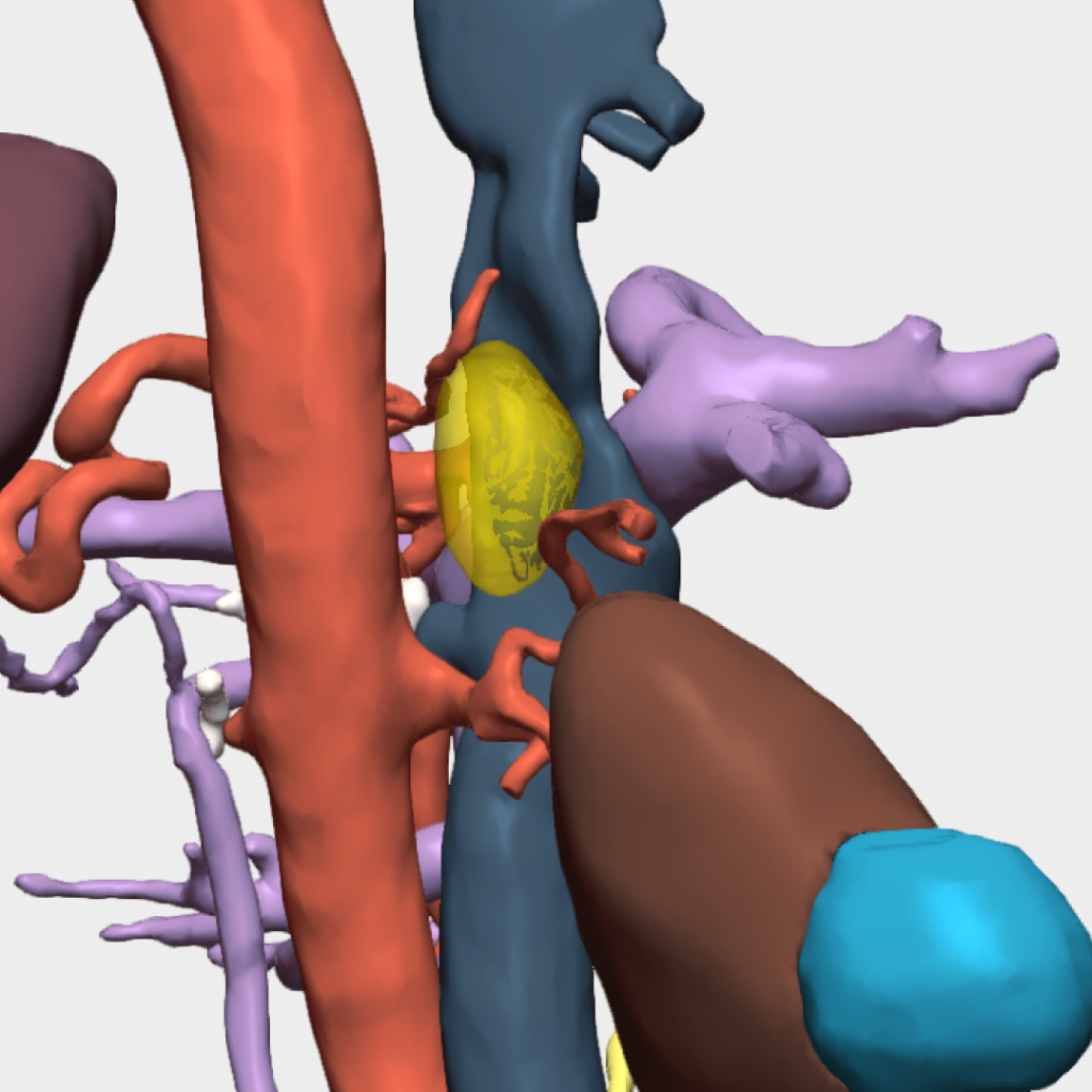
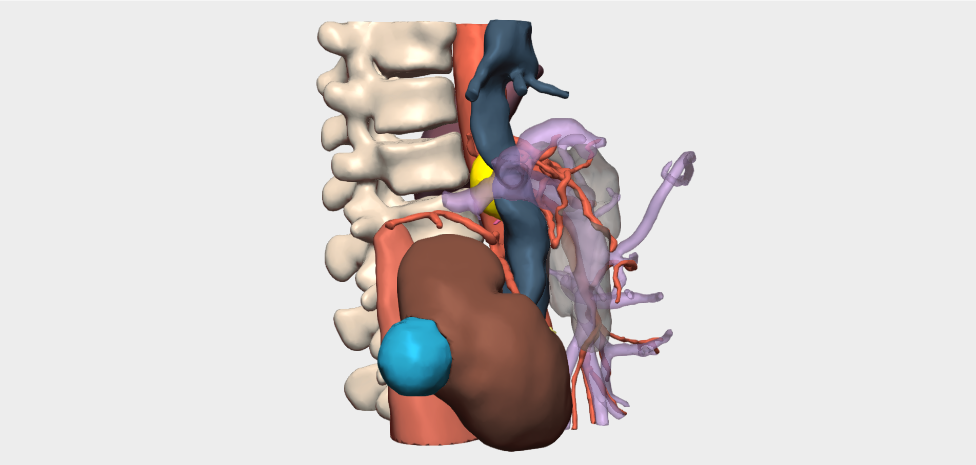
The 3D model proved highly valuable in analysing the tumour’s relationship with adjacent structures, confirming its intimate contact with the wall of the inferior vena cava and the right renal vein. This level of anatomical detail was essential for accurate risk assessment and preoperative strategy definition.
Based on the 3D findings, an open surgical approach via laparotomy was planned. The surgeons performed an extended Kocher manoeuvre to expose the retrohepatic and intrahepatic inferior vena cava, as well as the right renal vein, achieving optimal vascular control prior to tumour resection.
Surgical Outcome
Following five hours of surgery, and supported by meticulous preoperative planning and a clearly defined surgical strategy, complete excision of the paraganglioma was successfully achieved in this single-kidney patient.
The use of the 3D model contributed decisively to surgical precision, intraoperative safety and a fully planned operative approach, demonstrating its value in managing highly complex retroperitoneal tumours with major vascular involvement.
”The clinical value provided was extremely high, as the surgical field was anatomically complex and challenging to access. Thanks to the 3D model, the decision was made to proceed with an open surgical approach.
Dr María Lapeña & Dr. Norberto CassinelloHead of Section, Department of General Surgery and Consultant Oesophagogastric and Bariatric Surgeon