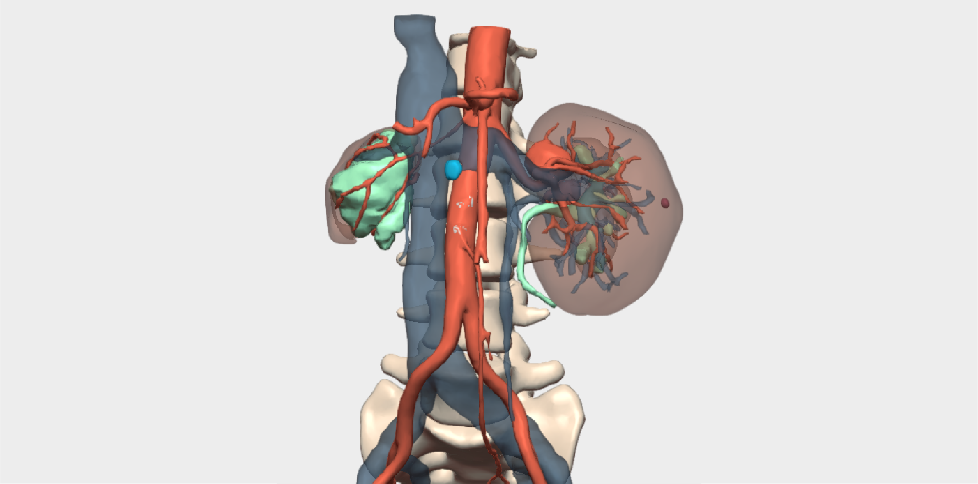

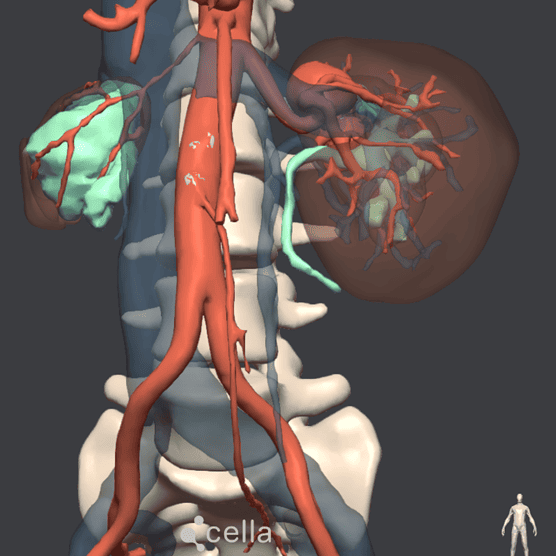
”Three-dimensional visualisation of the renal aneurysms and their close anatomical relationship with the renal parenchyma, vein and ureter was of key value in planning the repair to ensure the safest and most efficient surgical approach possible.
Dr. Francisco J. Gómez PalonésHead of the Vascular Surgery Department, Hospital Doctor Peset, Valencia
Los aneurismas en arteria renal son una dilatación anormal en los vasos que irrigan los riñones. Aunque poco frecuente, representa un riesgo significativo debido a la posibilidad de ruptura y al impacto sobre la función renal. En esta cirugía, realizada muy pocas veces en España, la planificación quirúrgica mediante una reconstrucción 3D fue clave para visualizar con precisión la anatomía compleja y definir una estrategia más segura y eficaz.
Clinical Case
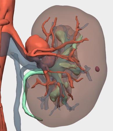
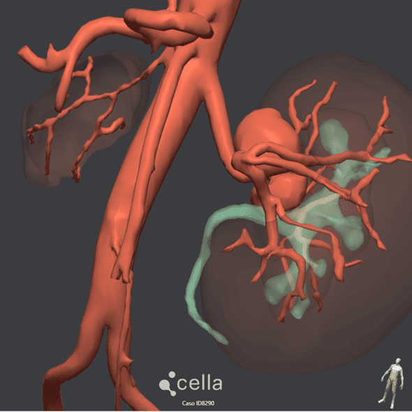
This case involves a 47-year-old female patient with a single functioning kidney, as the contralateral kidney had become non-functional due to underlying disease. The functioning kidney presented renal artery aneurysms in three different locations: within the main renal artery and in several of its branches extending into the renal hilum.
The aneurysmal dilatations had reached a considerable size, significantly increasing the risk of spontaneous rupture and haemorrhage, thereby posing a real, life-threatening risk to the patient. For these reasons, surgical repair of the aneurysms was strongly indicated.
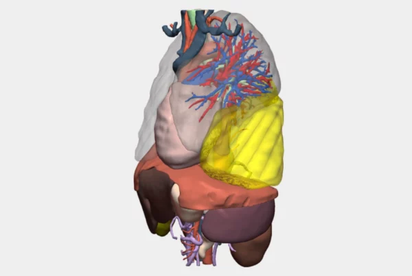
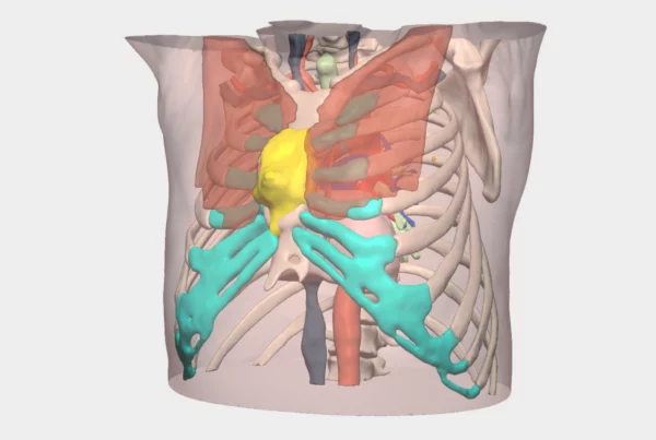
Renal Artery Aneurysms: Anatomical Complexity
The renal artery aneurysms were located not only very close to the renal hilum, but also involved multiple branch bifurcations, affecting tortuous arterial segments. This anatomical configuration considerably limited the feasibility of endovascular treatment options, such as embolisation.
While endovascular approaches may be appropriate in other anatomical settings, in this case the likelihood of preserving sufficient renal parenchyma was low, and the chances of achieving a durable long-term solution were even lower.
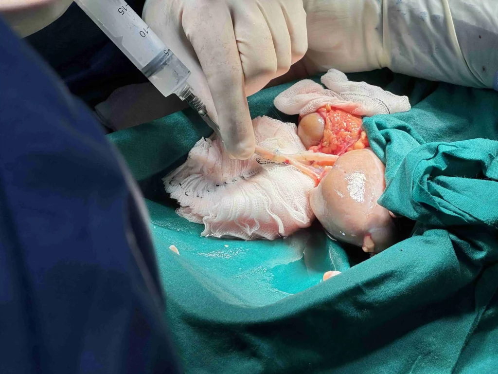
As a result, the most appropriate therapeutic option was ex vivo renal artery repair. This technique involves laparoscopic nephrectomy, followed by aneurysm repair outside the patient’s body. During the repair, the kidney is preserved using cold preservation solutions to prevent ischaemic damage during a reconstruction time that may exceed one hour. The kidney is then reimplanted in a more surgically favourable anatomical position, such as the right iliac fossa.
This approach is technically similar to the procedure used in living-donor kidney transplantation.
Value of the 3D Model in Surgical Planning
In this specific case, “3D visualisation of the renal aneurysms and their close anatomical relationships with the renal parenchyma, vein and ureter was of great value in planning the repair to ensure the safest and most efficient surgical approach possible,” explains Dr. Francisco J. Gómez Palonés, Head of Department and surgeon involved in the procedure.
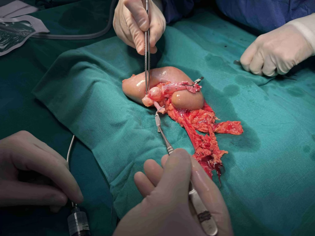
Renal aneurysm repair requires consideration of multiple surgical strategies, including venous interposition grafts harvested from other areas of the patient’s body, vessel transection and reimplantation, as well as lateral-to-lateral and end-to-side anastomoses at different arterial segments. The 3D model facilitated preoperative decision-making and strategy selection.
Surgical Outcome: Renal Artery Aneurysm Repair
The renal reconstruction procedure lasted 1 hour and 15 minutes. Thanks to meticulous preoperative planning supported by the 3D model, the surgical team successfully performed three arterial anastomoses between healthy arterial segments after aneurysm resection, avoiding the need to harvest venous grafts from the patient’s leg and preserving renal parenchyma.
The anatomical abnormality was fully corrected, and the patient’s renal function remained within normal limits, effectively eliminating the risk of aneurysm rupture.
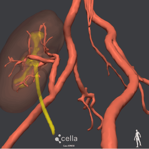
Postoperative Follow-Up and Assessment
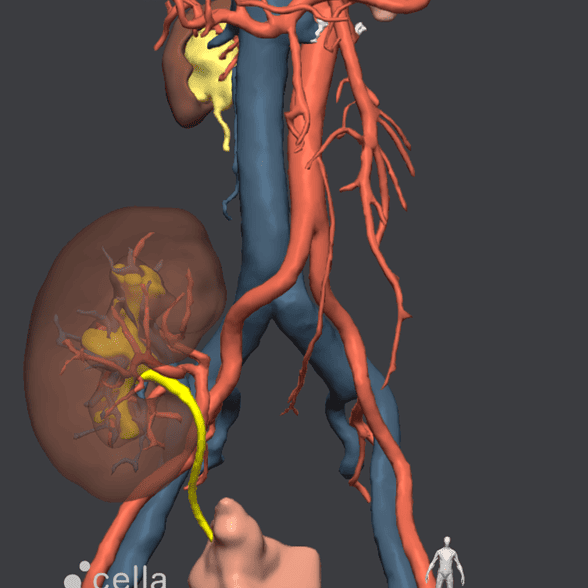
Following surgery and favourable clinical evolution, the surgical team requested a postoperative 3D model to analyse the reconstructed renal arterial anatomy in its new anatomical location, now free of aneurysms.
This postoperative 3D reconstruction enabled assessment of arterial patency and confirmation of the absence of new aneurysmal formations.
¡Follow us on social media to stay up to date with our latest clinical cases and technological innovations!