
”The decision was entirely appropriate, enabling the surgery to be completed successfully.
Dr Carolina Jiménez MazureGeneral and Digestive Surgeon, Complejo Hospitalario Regional de Málaga
Clinical Case
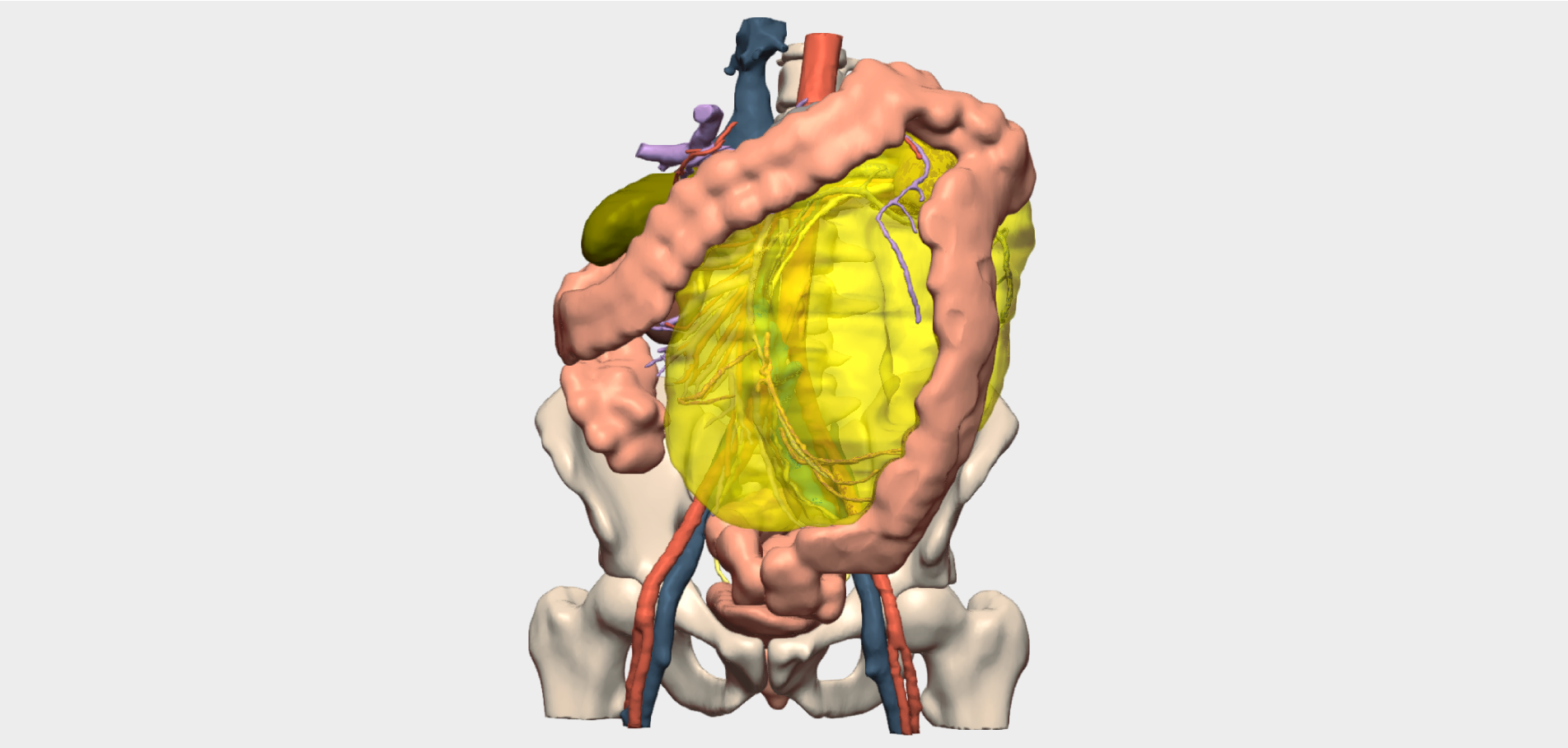
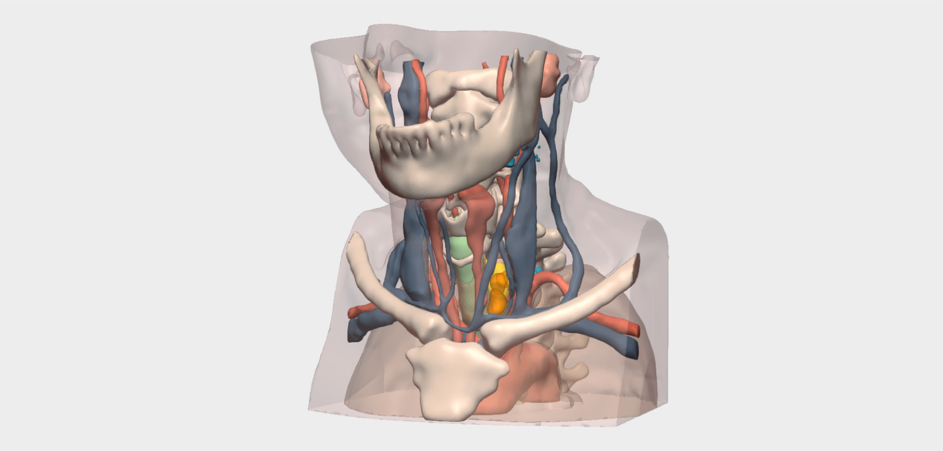
The patient presented with a parathyroid tumour associated with the inferior left thyroid lobe. Computed tomography (CT) imaging located the lesion in the lower third of the neck, in a complex anatomical position: posterior to the oesophagus with extension into the superior mediastinum.
This challenging location, together with the potential infiltration of adjacent structures, posed a significant surgical challenge. As a result, the surgical team requested a patient-specific 3D model to support preoperative planning and optimise the surgical approach.
Surgical Planning with the 3D Model
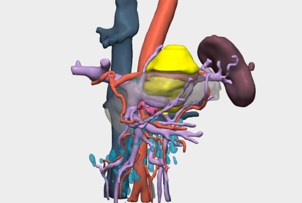
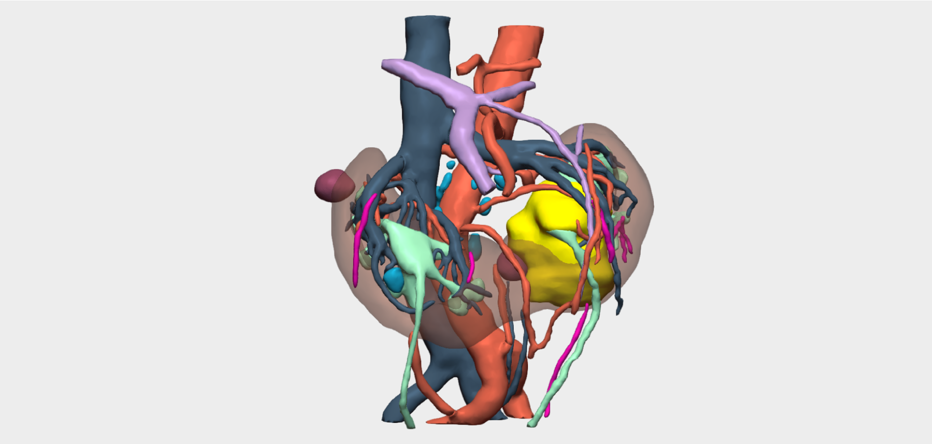
The use of the three-dimensional model was key to defining the surgical strategy. Advanced 3D functionalities enabled the surgical team to accurately visualise the tumour’s relationship with adjacent structures, including the oesophagus and the mediastinum.
Anatomical Relationship Analysis
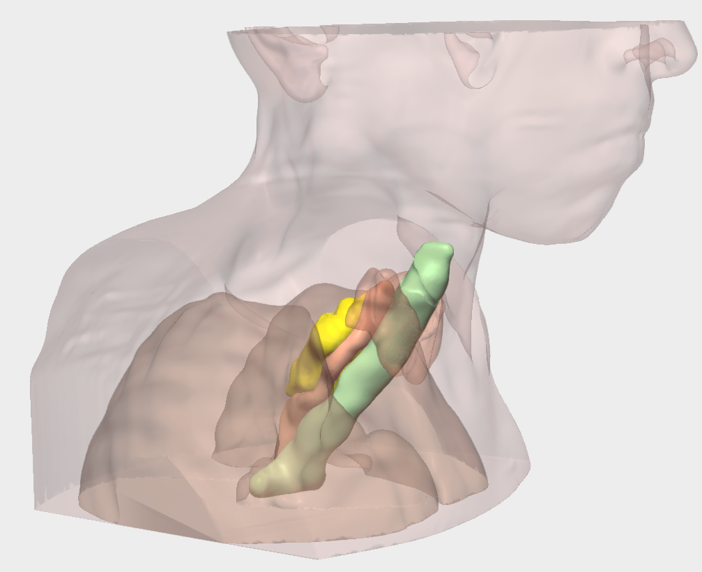
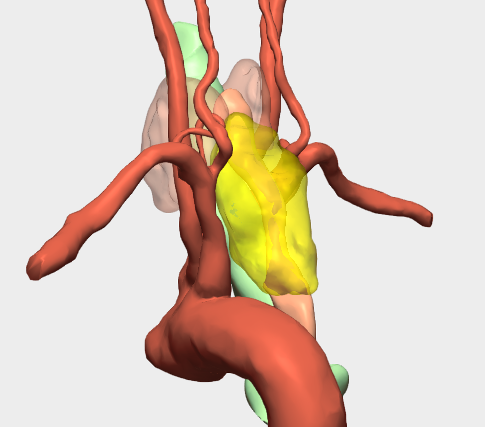
The three-dimensional model enabled precise analysis of the parathyroid mass and its relationship with adjacent structures, demonstrating excellent anatomical correlation between the cervical and intrathoracic components when assessed from all spatial perspectives.
Coordinated Intervention by Two Surgical Teams
Given the rarity of this tumoural pathology and the potential infiltration of adjacent structures, close coordination between multiple surgical teams was essential.
In addition to Dr Carolina Jiménez Mazure, Consultant General and Digestive Surgeon at Complejo Hospitalario Regional de Málaga, collaboration with Dr Roberto Mongil Ponce, Consultant Thoracic Surgeon at the same institution, was critical to the success of the procedure.
In this case, the 3D model enabled precise evaluation of the lesion’s location, size and anatomical relationships in both the cervical and thoracic regions, facilitating consensus between both teams and selection of the most appropriate surgical approach.
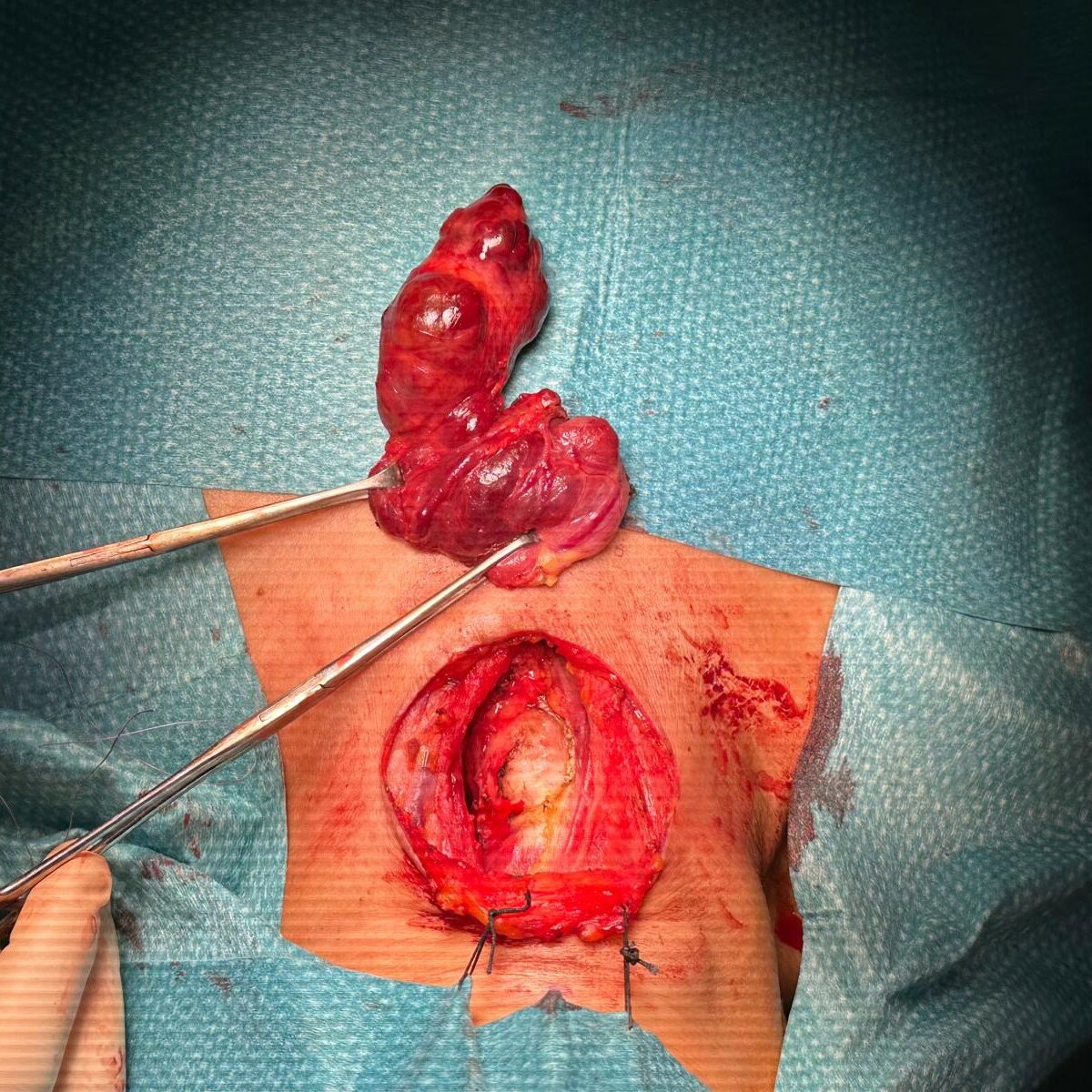
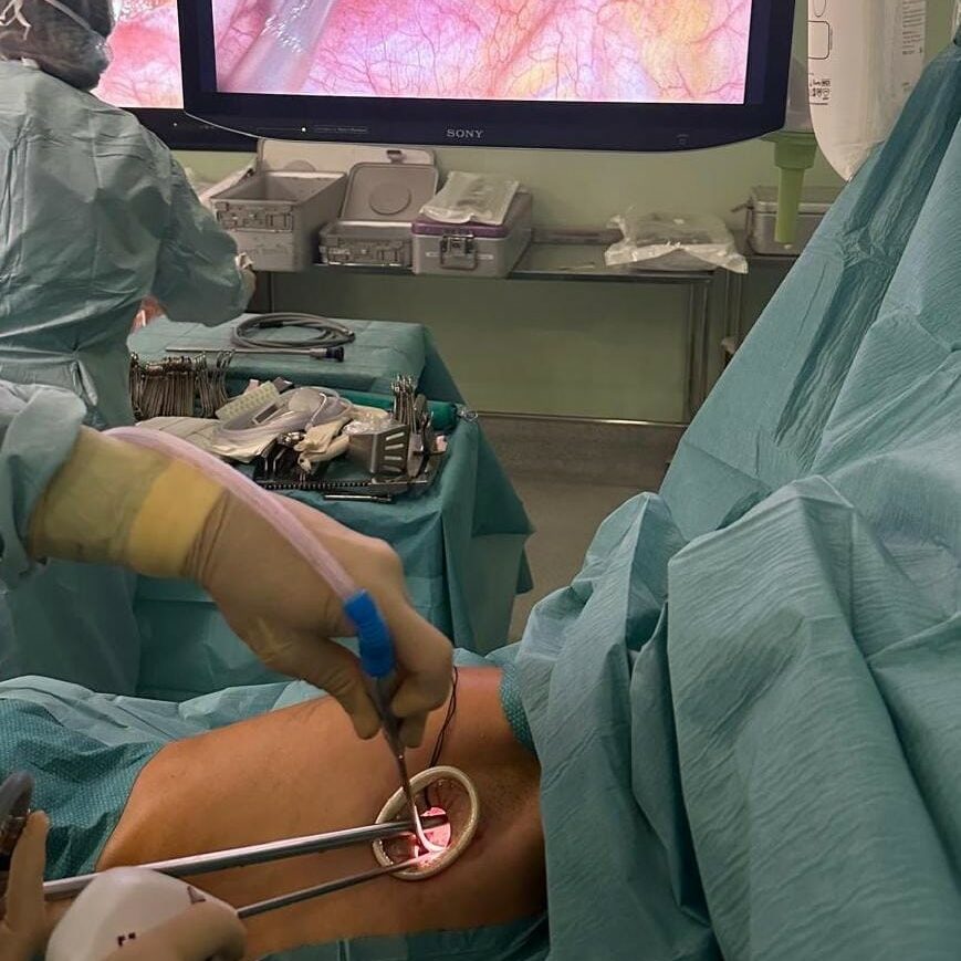
Surgical Outcome
This case highlights the value of 3D modelling as a transformative tool in surgical planning, particularly for highly complex procedures.
¡Follow us on social media to stay up to date with our latest clinical cases and technological innovations!