”The model helped us to orient ourselves following the initial surgery performed in Morocco and also alerted us to the potential risk of injury to the vascular and bronchial structures of the small remaining lung tissue
Dr. Óscar GirónHead of Section of Paediatric Surgery, Hospital Universitario Virgen de la Arrixaca
Clinical Case
We present the case of a 13-year-old female patient, originally from Morocco, with recurrent CPAM (Congenital Pulmonary Airway Malformation) affecting the right lung. The patient had undergone surgery in her country of origin at three months of age and, thirteen years later, required reintervention at Hospital Universitario Virgen de la Arrixaca.
One of the main challenges in this case was the discrepancy between the information available regarding the initial surgery performed in Morocco and the findings observed on the current CT scan, which introduced significant uncertainty into preoperative planning.
3D Model–Assisted Surgical Planning
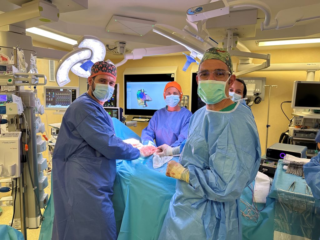
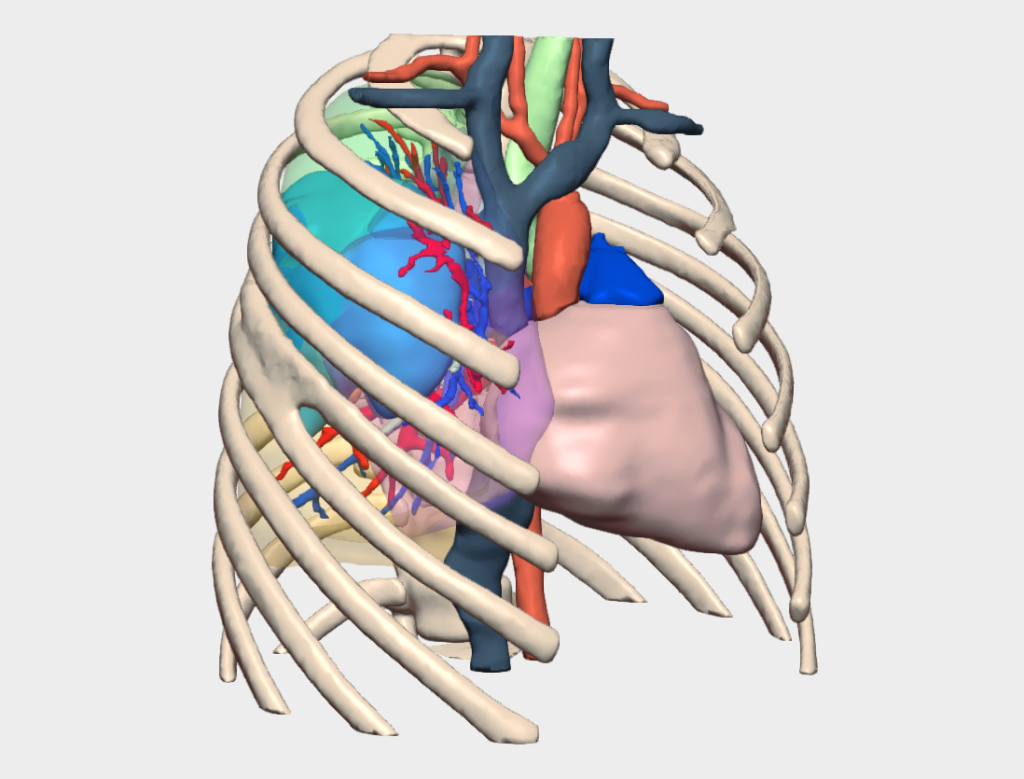
Given the complexity of the case, the surgical team decided to complement preoperative planning with a patient-specific 3D model.
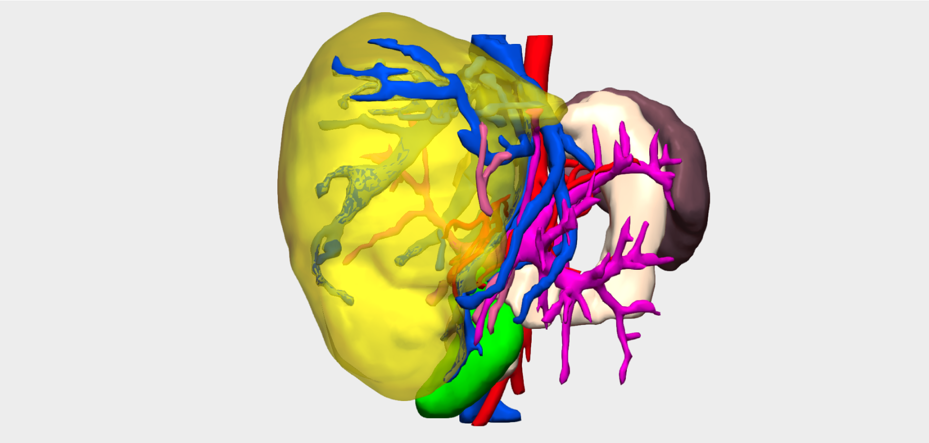
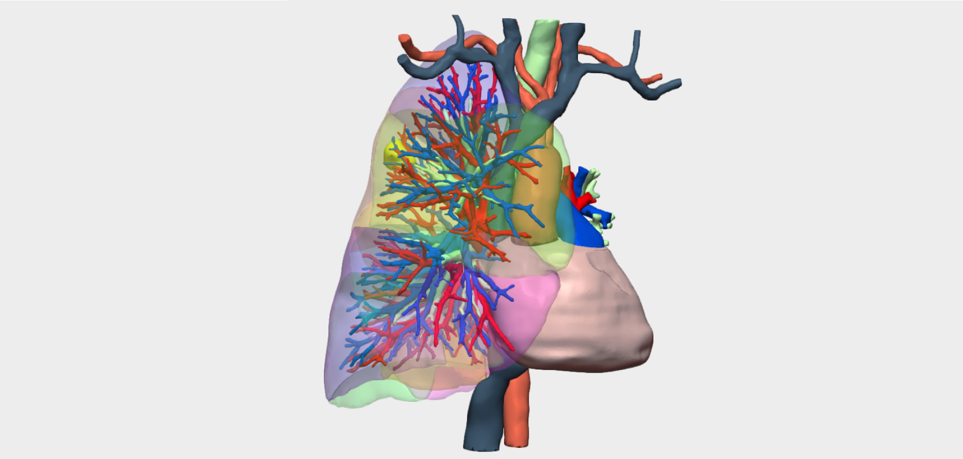
“The model helped us orient ourselves following the initial surgery performed in Morocco and also alerted us to the potential risk of injury to vascular and bronchial structures of the small remaining lung tissue,” explains Dr. Óscar Girón, Head of Section of Paediatric Surgery at Hospital Universitario Virgen de la Arrixaca.
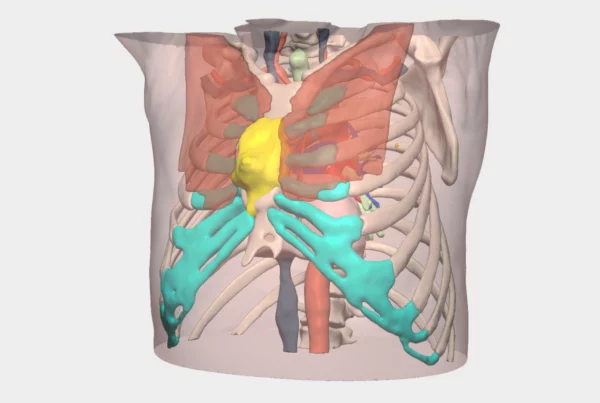
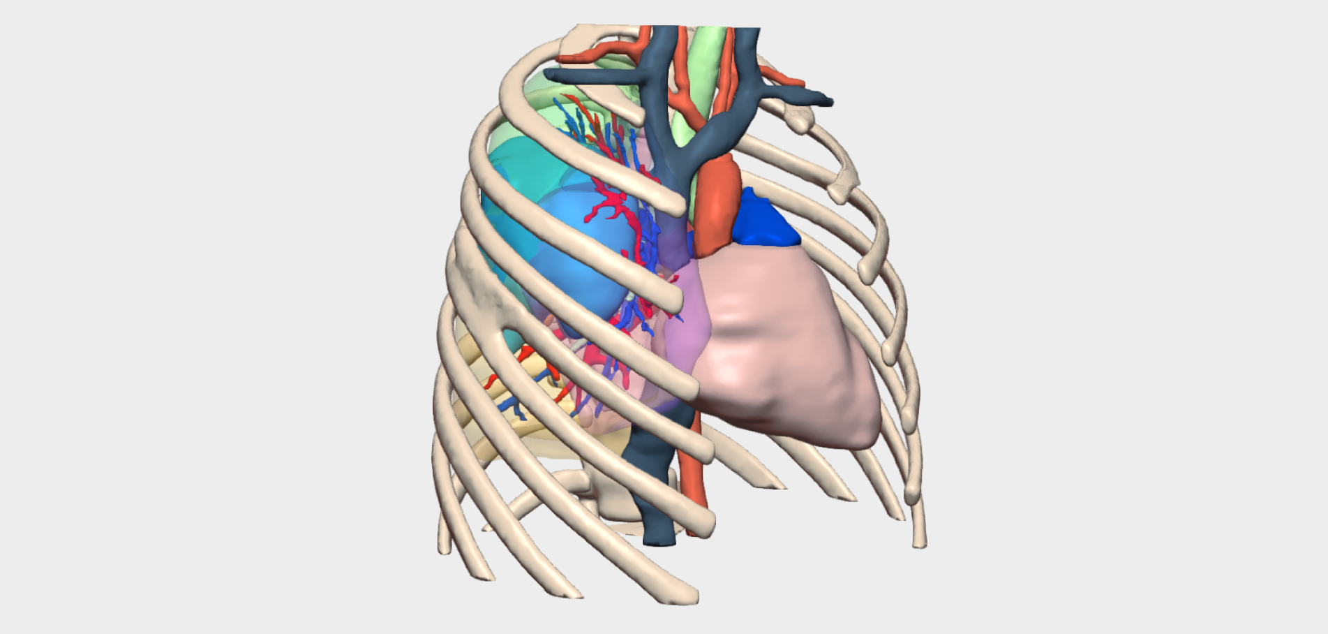
In addition, the 3D model enabled clear visualisation of rib fusion resulting from the previous intervention, allowing the team to plan the most appropriate intercostal access to the malformation.
The surgical strategy adopted by the multidisciplinary team at Hospital Universitario Virgen de la Arrixaca—comprising Dr Ramón Ruiz, Dr Óscar Girón, and the Head of the Thoracic Surgery Department—consisted of a cystectomy through a non-anatomical segmentectomy, tailored to the specific anatomical conditions of the patient. The primary objective was to avoid right pneumonectomy and preserve as much functional lung tissue as possible.
Surgical Outcome
The patient remained hospitalised for 10 days and was discharged without complications, with adequate re-expansion of the remaining lung tissue. In this highly complex case, the use of a 3D model proved to be decisive in avoiding a potential right pneumonectomy, enabling preservation of the lung with good functional tolerance and a favourable clinical outcome