Next Generation
3D modelling
SUCCESS STORIE
Dr. Iván Bautista
Hospital Teletón Infantil de Oncología
Clinical Case
Patient-Specific 3D Model of a Paediatric Liver Tumour | Cella Medical Solutions
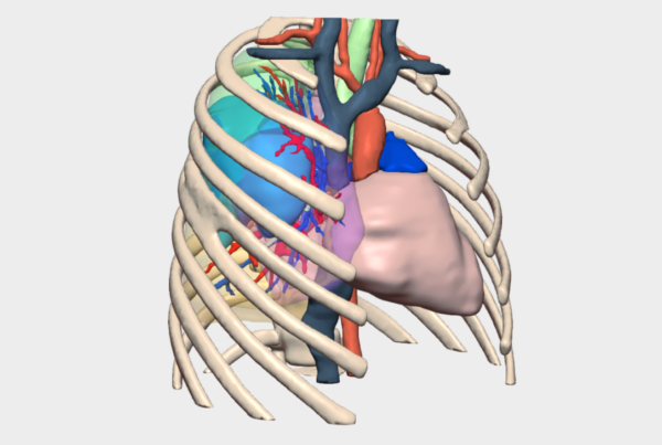
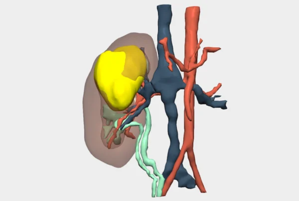
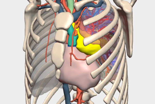
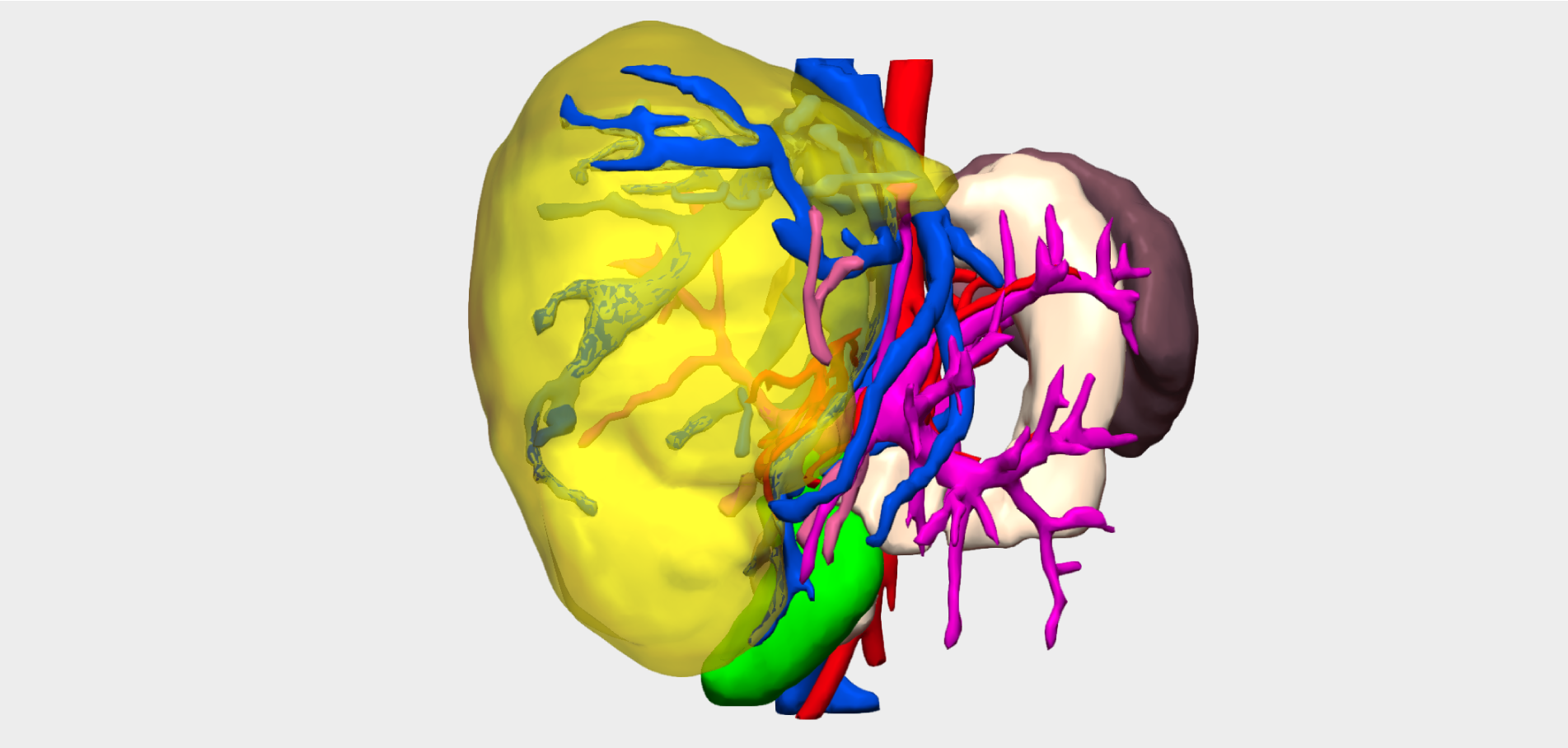
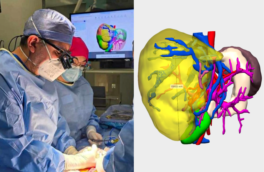
We present the case of a paediatric patient with a highly complex liver tumour, characterised by its large size and involvement of major vascular structures.
Value of the 3D Model in Surgery
Thanks to the patient-specific 3D model, Dr Iván Bautista, Consultant Paediatric Surgeon at the Hospital Teletón Infantil de Oncología (Fundación Teletón), Mexico, obtained a detailed visualisation of the hepatic anatomy and was able to accurately assess the implications of the lesion, which was shown on the 3D model to be of considerable size.
The 3D model was particularly valuable for visualising the hepatoduodenal ligament and understanding the course of the hepatic artery. This information was critical for the surgical team in precisely planning tumour resection in segment II.
Surgical Outcomes
Following a six-hour procedure, the hospital’s surgical team achieved complete resection of a 1.9 kg tumour through an extended right hepatectomy, involving removal of segments IV, V, VI, VII and VIII.
During surgery, a 45-minute Pringle manoeuvre was performed, with a total estimated blood loss of 500 ml.
Postoperative pathological analysis confirmed an unclassified tumour, consisting of a mixed hepatoblastoma and hepatocellular carcinoma, the latter being extremely rare in paediatric patients.
Three-dimensional planning and visualisation were decisive to the success of this highly complex paediatric liver surgery, enabling accurate surgical planning and effective tumour resection while optimising intraoperative safety.
”The anatomy of the hepatoduodenal ligament showed us the course of the artery and was extremely helpful during dissection of the segment II tumour.
Dr. Iván BautistaConsultant Paediatric Surgeon, Hospital Teletón Infantil de Oncología