“3D reconstruction represents a highly valuable tool, providing detailed anatomical information and spatial visualization that facilitates surgical decision-making and reduces intraoperative risks.”

Dr. Manuel Abradelo de Usera, Lead Surgeon, Toledo University Hospital Complex
Case Presentation
A 48-year-old female patient with no significant past medical history presented with poorly controlled arterial hypertension and nonspecific neurological symptoms (headaches, orthostatic hypotension, and presyncope). She was referred to the Toledo University Hospital Complex for further evaluation.
Abdominopelvic CT scan revealed a large left retroperitoneal mass (7.5 cm in maximum diameter), in close contact with the left adrenal gland, splenic artery and vein, and the pancreatic tail, with infiltration not ruled out. Imaging findings were suggestive of pheochromocytoma/paraganglioma.
Surgical Challenges
The case involved high surgical complexity due to:
- Uncertain origin of the retroperitoneal lesion.
- Possible pancreatic infiltration, potentially requiring pancreatectomy with associated risks (hemorrhage, pancreatic fistula).
- Suspected vascular involvement, which could necessitate splenectomy and complex vascular reconstruction.
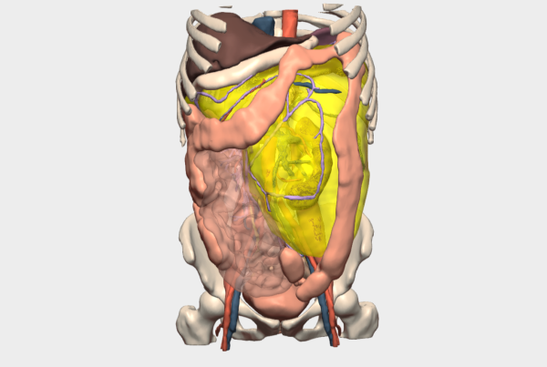
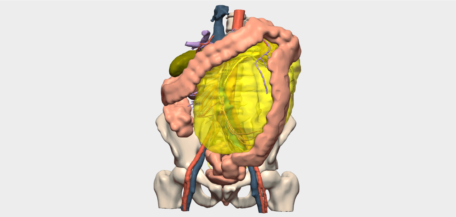
Given the need to achieve a radical (R0) resection and the significant technical risk, a 3D model was used to accurately assess the tumor’s relationship with vascular structures and adjacent organs, enabling safer and more precise surgical planning.
3D Model–Based Surgical Planning
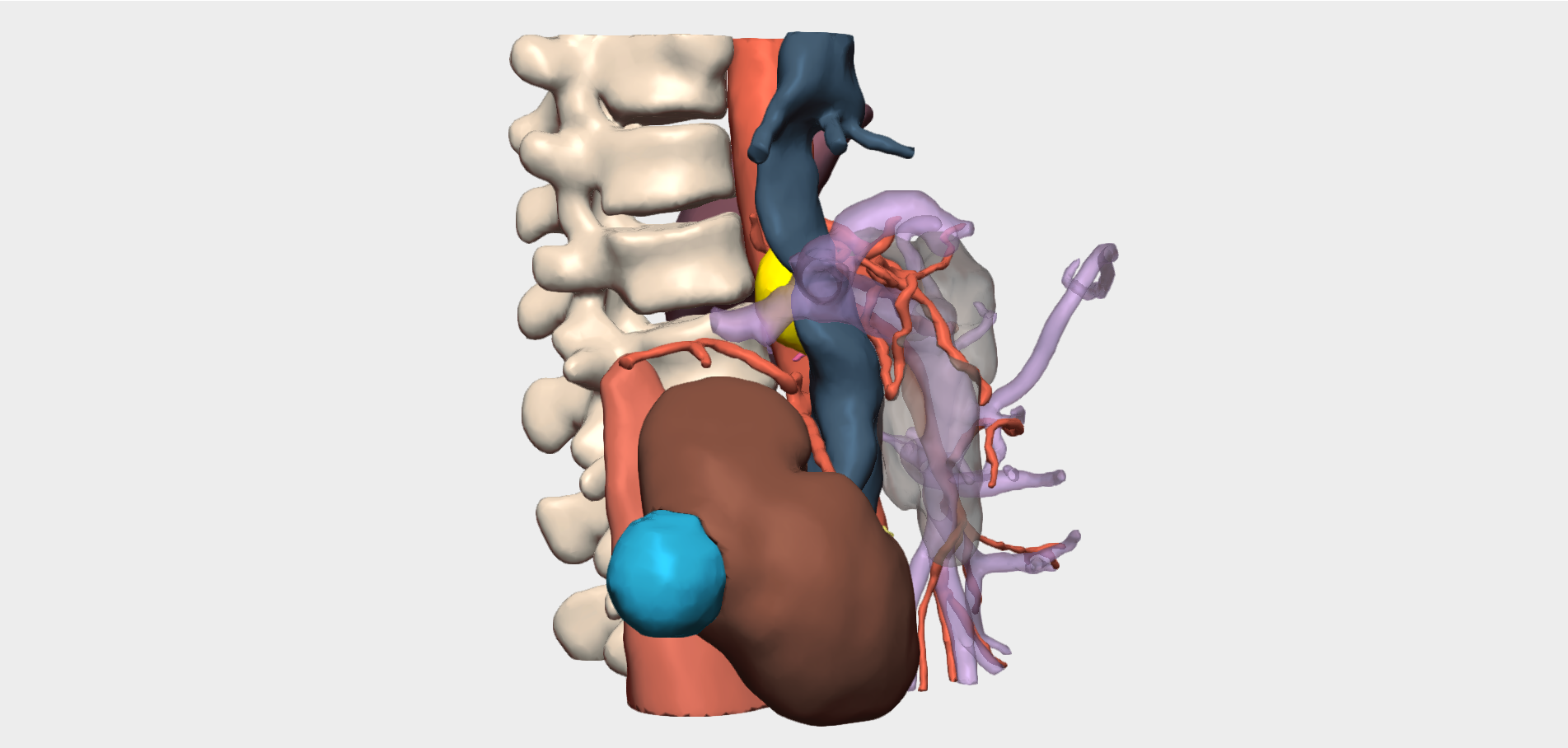
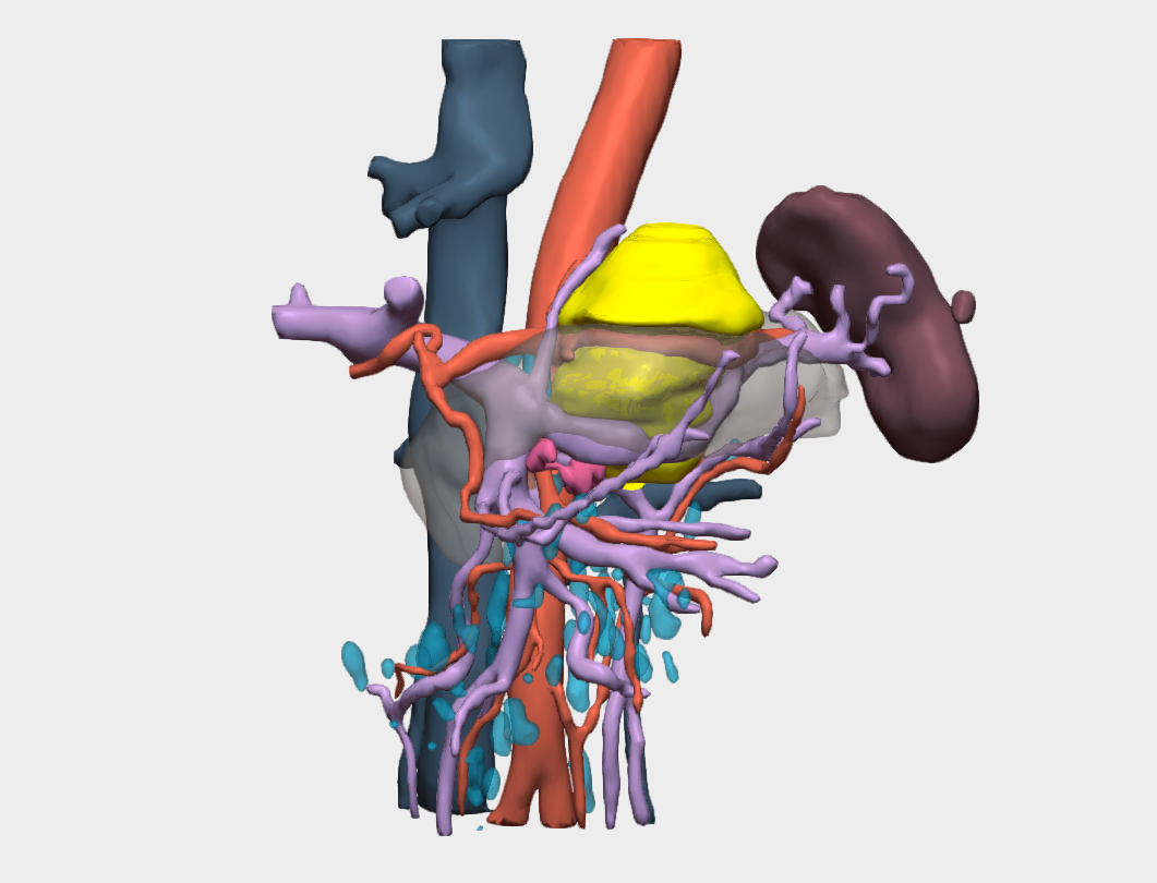
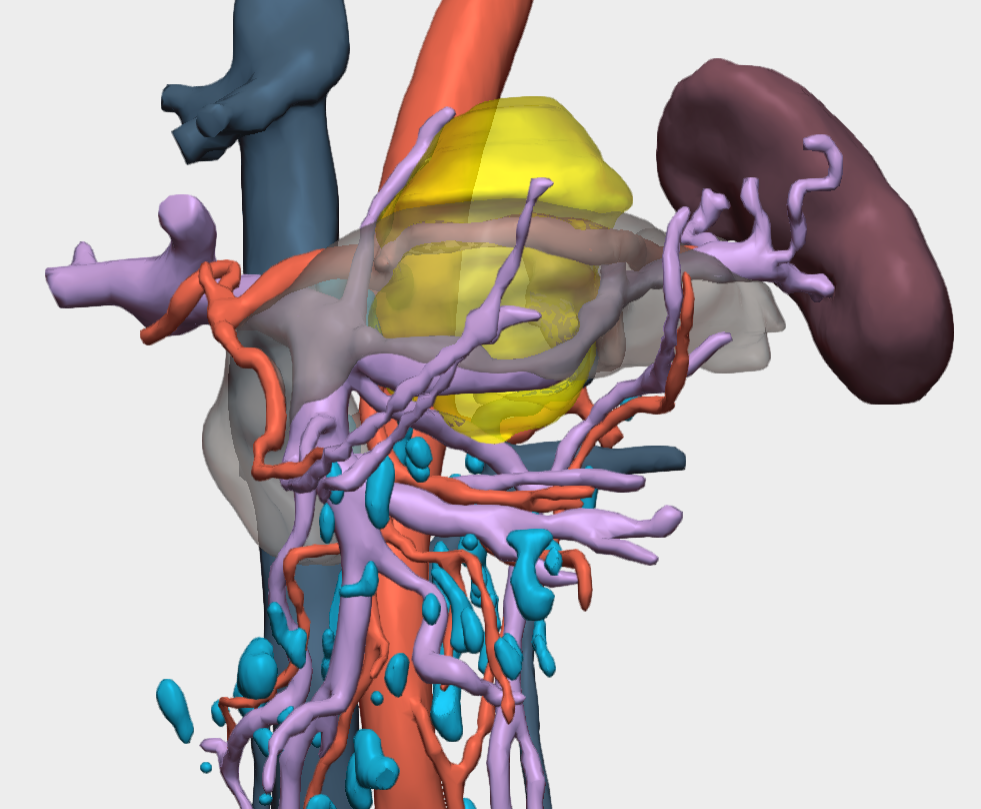
The 3D reconstruction enabled precise isolation and analysis of key structures: pancreas, splenic vessels, adrenal gland, and mesocolon. The model confirmed:
- Involvement of the splenic artery at its origin, with partial extension to the celiac trunk.
- Partial involvement of the mesocolon and intimate contact with the pancreas and adrenal gland.
This advanced visualization facilitated strategic surgical decision-making, refining the operative approach and enhancing team coordination.
Surgical Procedure and Strategy
The surgical intervention included:
- Open (laparotomic) resection of the retroperitoneal mass.
- Distal pancreatectomy with splenectomy.
- Left adrenalectomy.
- Resection of a mesocolic patch (intraoperative biopsy ruled out malignancy).
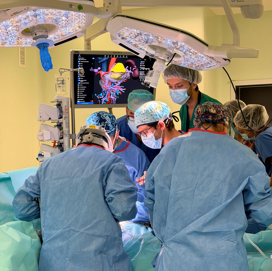
The procedure was performed by Dr. Manuel Abradelo de Usera as lead surgeon, assisted by Dr. David Martínez Cecilia, Dr. Luis Cadaval, and Dr. Lucas Rodríguez-Carreño Díaz, with collaboration from Dr. Javier Lesaga Llopis.
Outcomes and Conclusions
The surgery was completed without complications, achieving controlled dissection and complete tumor resection (R0). The patient was admitted to the Intensive Care Unit for 24 hours for hemodynamic monitoring and was discharged on postoperative day six with favorable clinical evolution.
This case highlights how advanced surgical planning using 3D models provides detailed anatomical insight, anticipates risks, and enhances surgical safety. In complex retroperitoneal tumors, where the relationship with major vessels and adjacent organs is critical, three-dimensional reconstruction becomes an indispensable tool for optimal clinical outcomes.
¡Follow Us!