Next Generation
3D modelling
Success Storie
Dr. Alejandro García-Seguí
Hospital General Universitario de Elche
Clinical Case
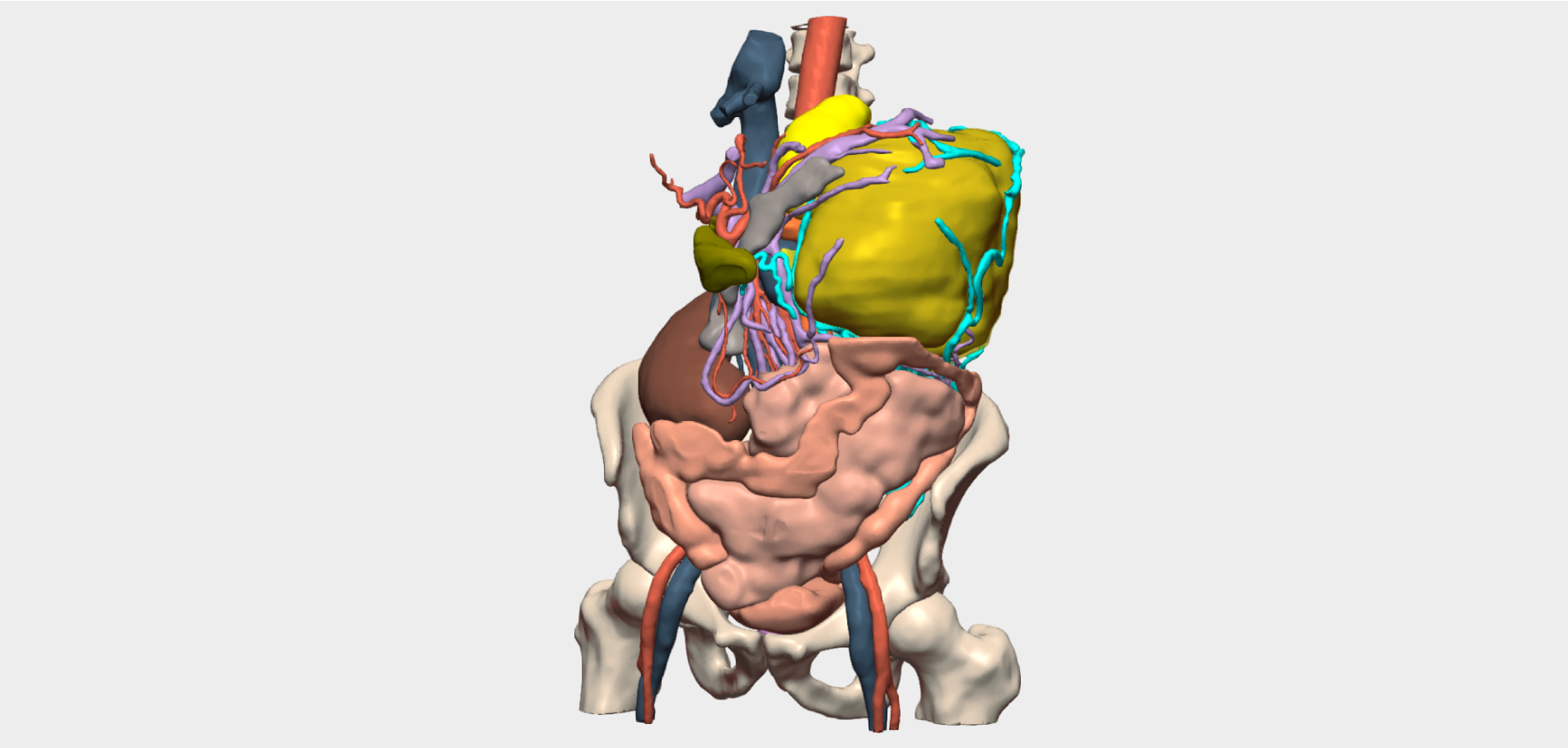
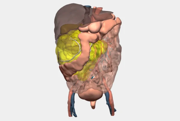
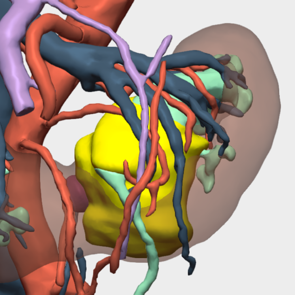
Patient-Specific 3D Model of a Horseshoe Kidney – Cella Medical Solutions
This case involves an 86-year-old male patient diagnosed with a horseshoe kidney presenting a Type VI segmental arterial pattern according to the Graves classification. The patient was receiving anticoagulant therapy due to atrial fibrillation, further increasing surgical complexity.
A large urothelial tumour of the left renal pelvis was identified, and a laparoscopic left hemi-nephroureterectomy was planned.
Value of the 3D Model in Surgical Planning
Dr Alejandro García-Seguí, Consultant Urological Surgeon and lead surgeon for this procedure, supported surgical planning and execution using a patient-specific 3D model, highlighting its value for the following reasons:
-
-
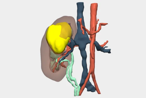
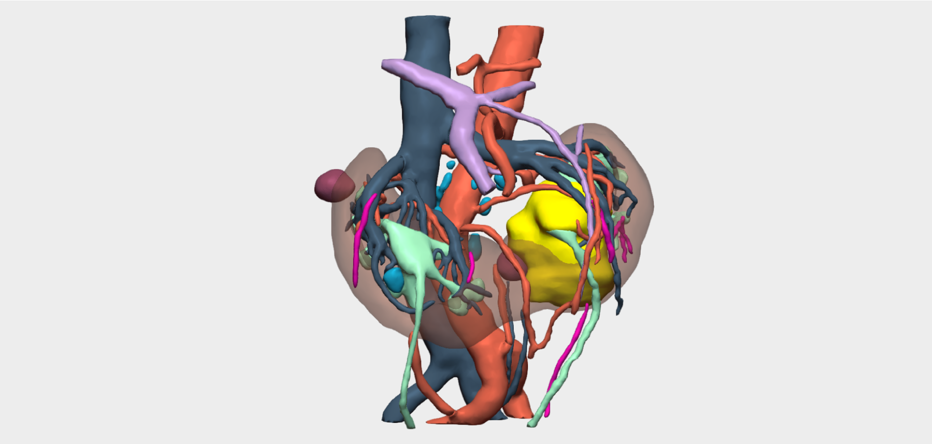
It facilitated targeted planning of the surgical approach, enabling identification of all arterial branches supplying the affected renal moiety, as well as control of surrounding arteries at the renal isthmus during renal transection and renorrhaphy.
-
It ensured isthmus transection with negative surgical margins by using a renal cyst as an anatomical reference point, allowing precise localisation of the renal tumour.
-
It confirmed preservation of the lower calyceal system of the contralateral renal moiety during isthmus section, which was infundibulised and located in close proximity to the tumour.
-
Surgical Strategy
The surgical approach implemented by the medical team included:
-
Initial identification of the left ureter
-
Dissection of the external surface of the aorta
-
Systematic identification—supported by the 3D model—of all arterial vessels supplying the left renal moiety, totalling six arteries
-
Ligation of the complex left renal vein
-
Dissection of the renal isthmus and associated vessels
-
Isthmus transection using thermal energy followed by two-layer renorrhaphy
Surgical Outcome
Following a 200-minute procedure, surgery was completed successfully, achieving negative surgical margins, minimal intraoperative blood loss, and no postoperative anaemia or haemorrhagic complications.
”The 3D model was extremely useful, as it enabled a targeted surgical approach focused on identifying the vascular supply of the affected renal moiety, ensuring isthmus transection with negative surgical margins.
Dr. Alejandro García-SeguíConsultant Urologist specialised in Laparoscopic Surgery, Hospital General Universitario de Elche