“The 3D model proved extremely valuable in planning the surgical approach to the vascular pedicle and enabling tumour resection while preserving adjacent organs and major vessels, particularly the aorta and the inferior vena cava”
Dr. Ignacio Rubio Tortosa, Urological Surgeon, Hospital Virgen de los Lirios, Alicante
Clinical Case
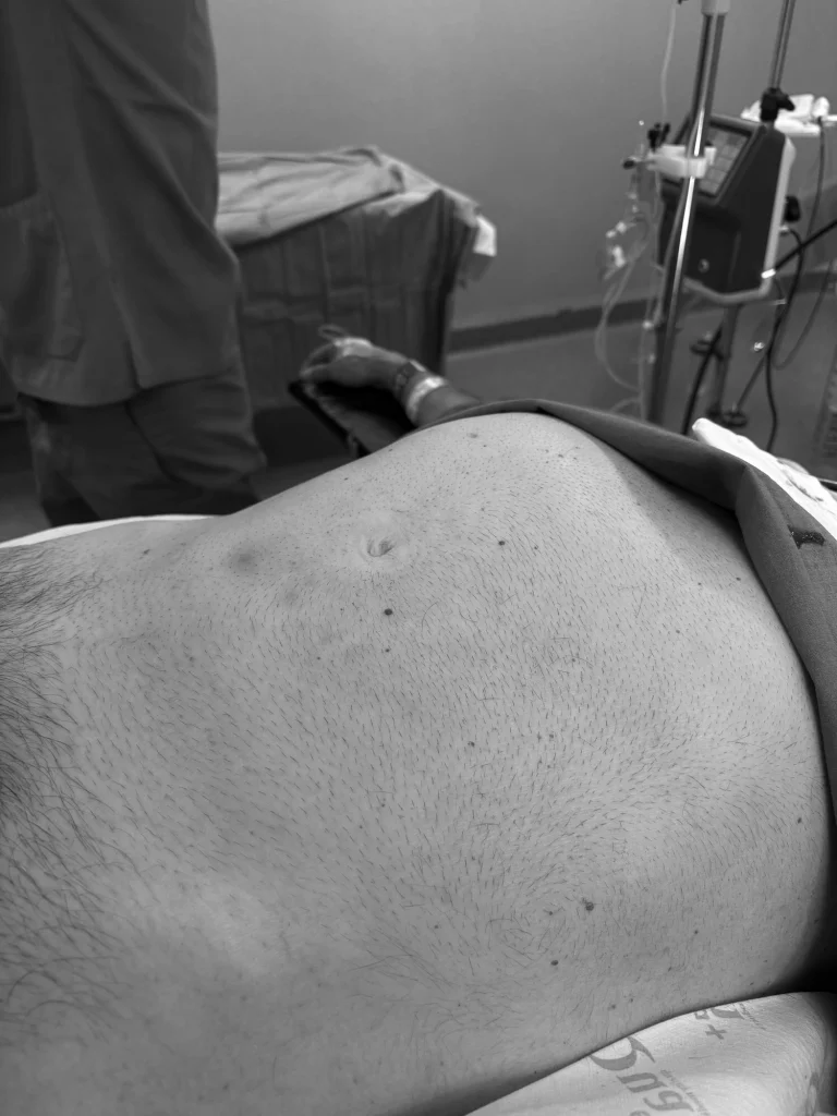
This case involves a 64-year-old male patient with no relevant medical history, who consulted his General Practitioner due to progressive enlargement of the right hemiabdomen over several weeks, without associated symptoms.
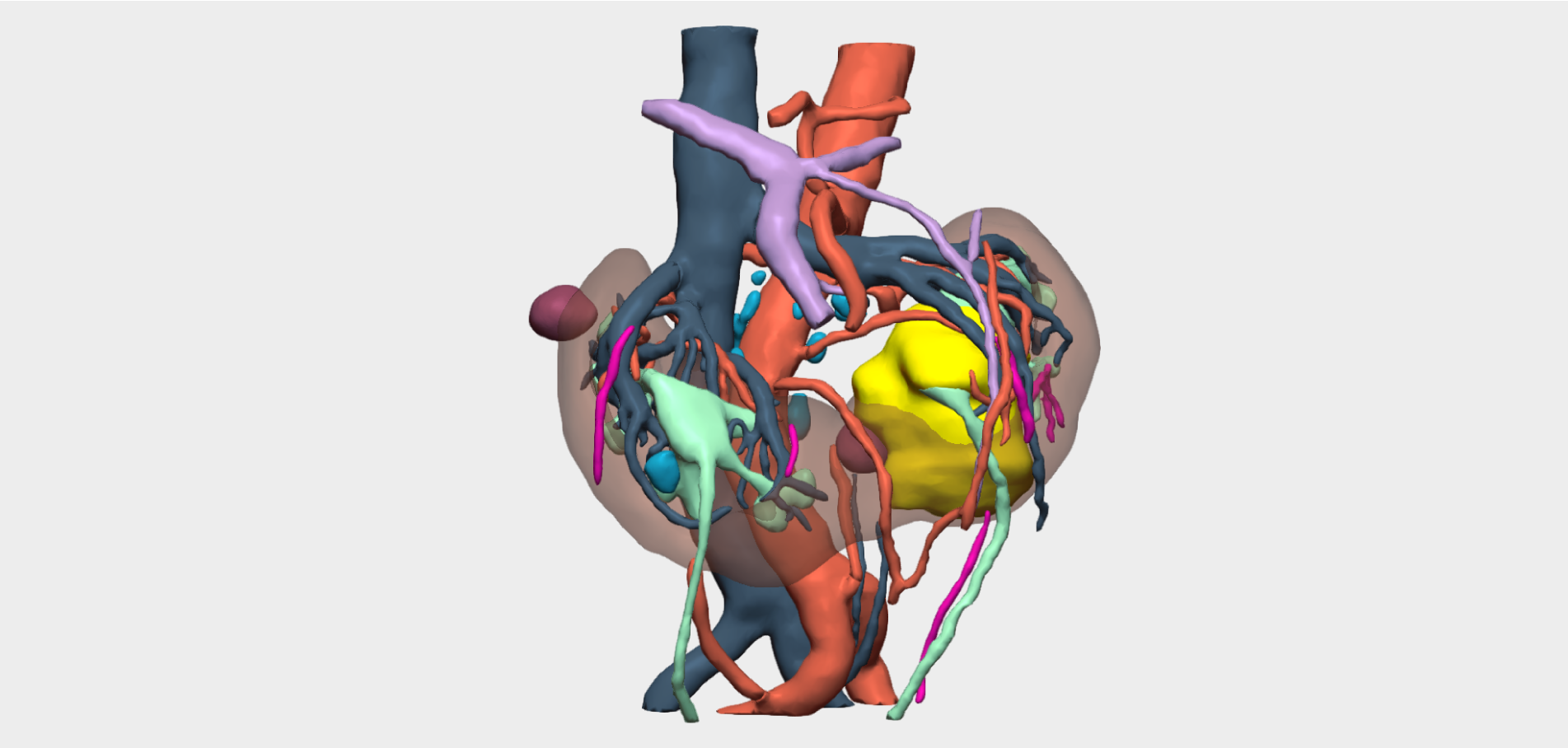
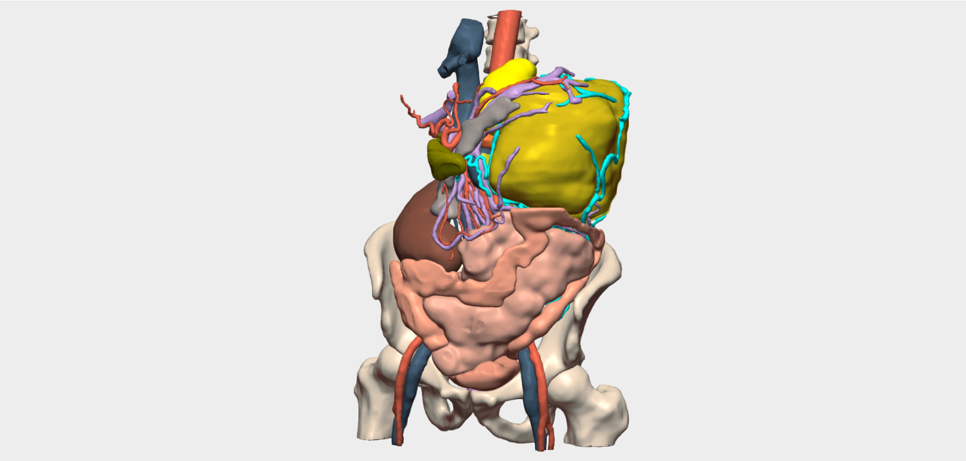
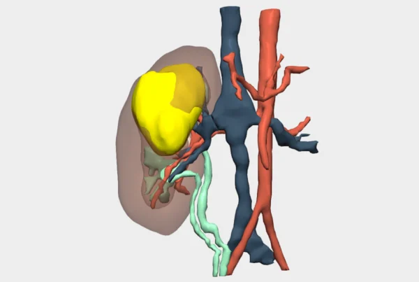
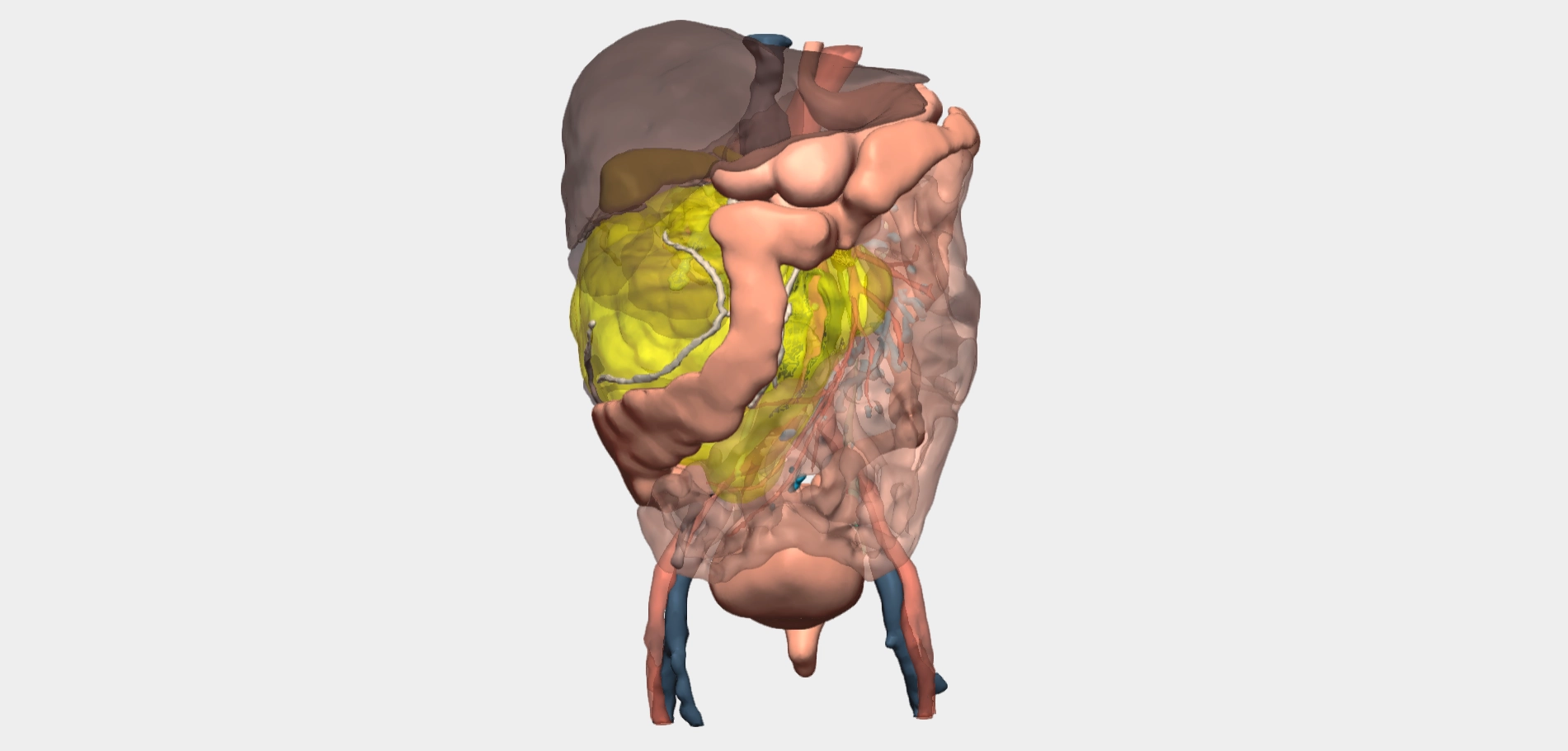
Ultrasound and computed tomography (CT) imaging revealed a giant tumour arising from the right kidney. The mass displaced the intestinal loops and extended almost to the spleen and the bladder. No lymphadenopathy or distant metastases were identified. The tumour reached a size sufficient to displace the aorta and cause near-complete compression of the inferior vena cava.
3D Planning of High-Volume Renal Tumour Surgery
In light of these findings, the patient was referred to the Urology Department at Hospital Universitario Virgen de los Lirios in Alcoy (Alicante). A patient-specific 3D study was requested from Cella Medical Solutions to support radical surgical planning, with particular focus on neighbouring organs and, critically, the involvement of major vascular structures.
Given the complexity of the case, the reference Vascular Surgery Department within the regional Health Authority was consulted but declined participation or joint intervention. Similarly, the General Surgery Department at the same hospital was asked to assess potential involvement; however, collaboration was also declined due to the technical difficulty of the procedure.
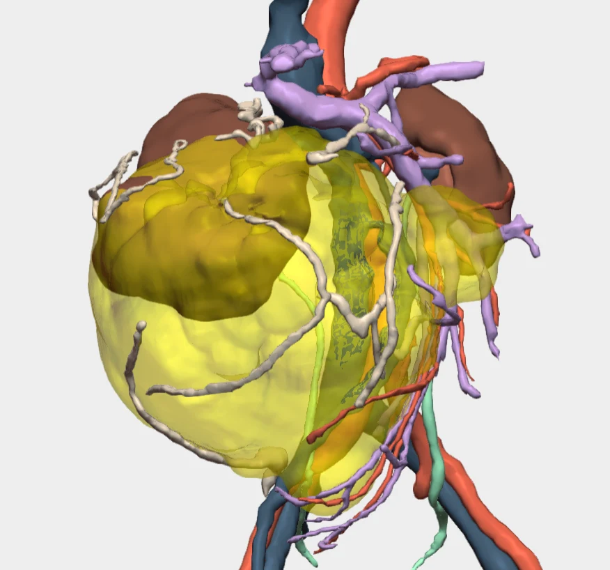
Ultimately, the surgery was performed exclusively by the Urology Department at Hospital Virgen de los Lirios, with Dr Ignacio Rubio Tortosa as lead surgeon. The 3D model played a key role in defining the optimal approach to the vascular pedicle and in enabling complete tumour resection while preserving adjacent organs and major vessels, particularly the aorta and the inferior vena cava.
Surgical Outcome
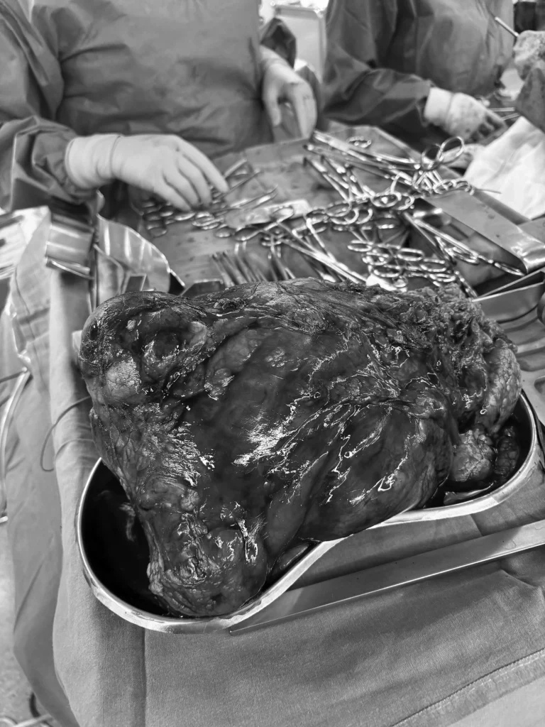
Despite the high technical complexity of the procedure, complete tumour resection was achieved without immediate complications, and the patient experienced early postoperative recovery.
Histopathological analysis of the surgical specimen confirmed a 21 cm chromophobe renal cell carcinoma with invasion of the renal sinus fat (pT3aN0).
The case was subsequently reviewed by the multidisciplinary tumour board, where adjuvant treatment was deemed unnecessary. The patient is currently in complete remission and in good general condition.
Follow us on social media to stay up to date with our latest clinical cases and innovations.