Next Generation
3D modelling
SUCCESS STORY
Dra. Yolanda Quijano
Hospital Universitario HM Sanchinarro
Clinical Case
3D Surgical Planning
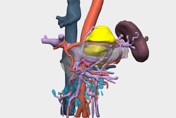
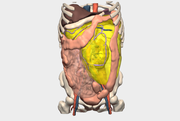
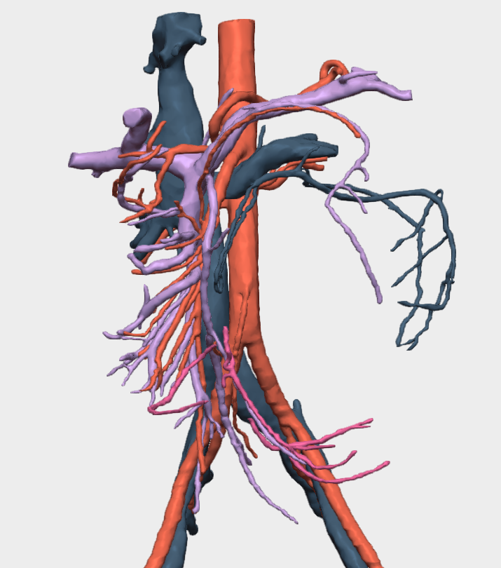
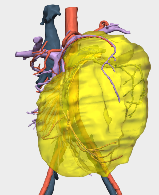
To support preoperative planning, the hospital’s medical team utilised a patient-specific 3D model of the patient’s anatomy. Analysis of the model revealed that the lesion caused significant displacement of mesenteric structures towards the right side, externally and posteriorly displacing the descending colon.
In addition, vascular structures were also involved in this displacement, adopting the contour and morphology of the tumour, a critical factor for surgical risk assessment and planning.
Surgical Approach
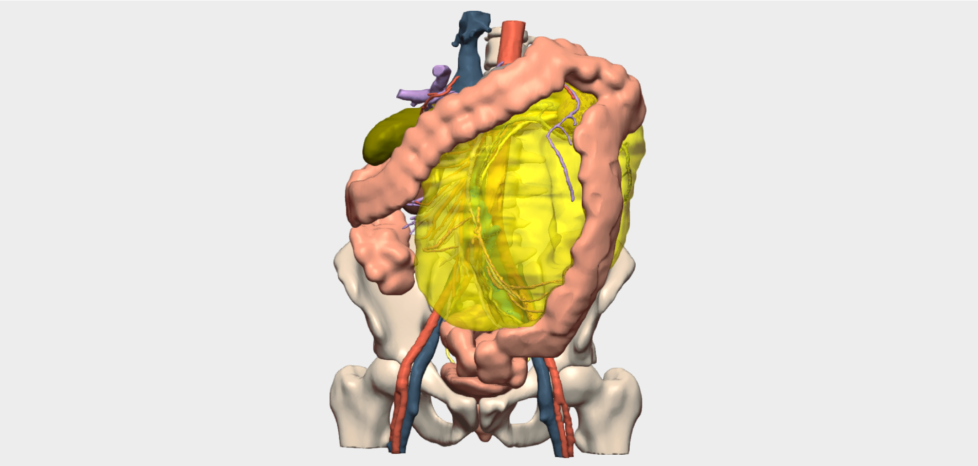
During surgery, the team successfully preserved the left colon, an outcome that initially appeared unlikely based on conventional preoperative CT imaging alone. This preservation was made possible by the detailed anatomical visualisation provided by the 3D model.
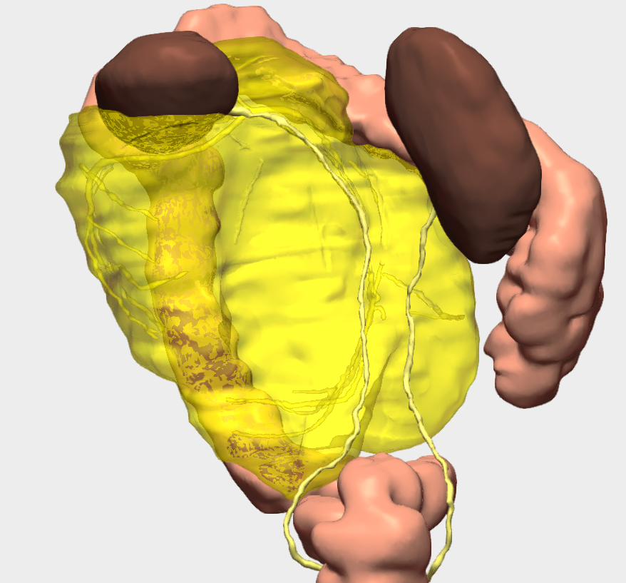
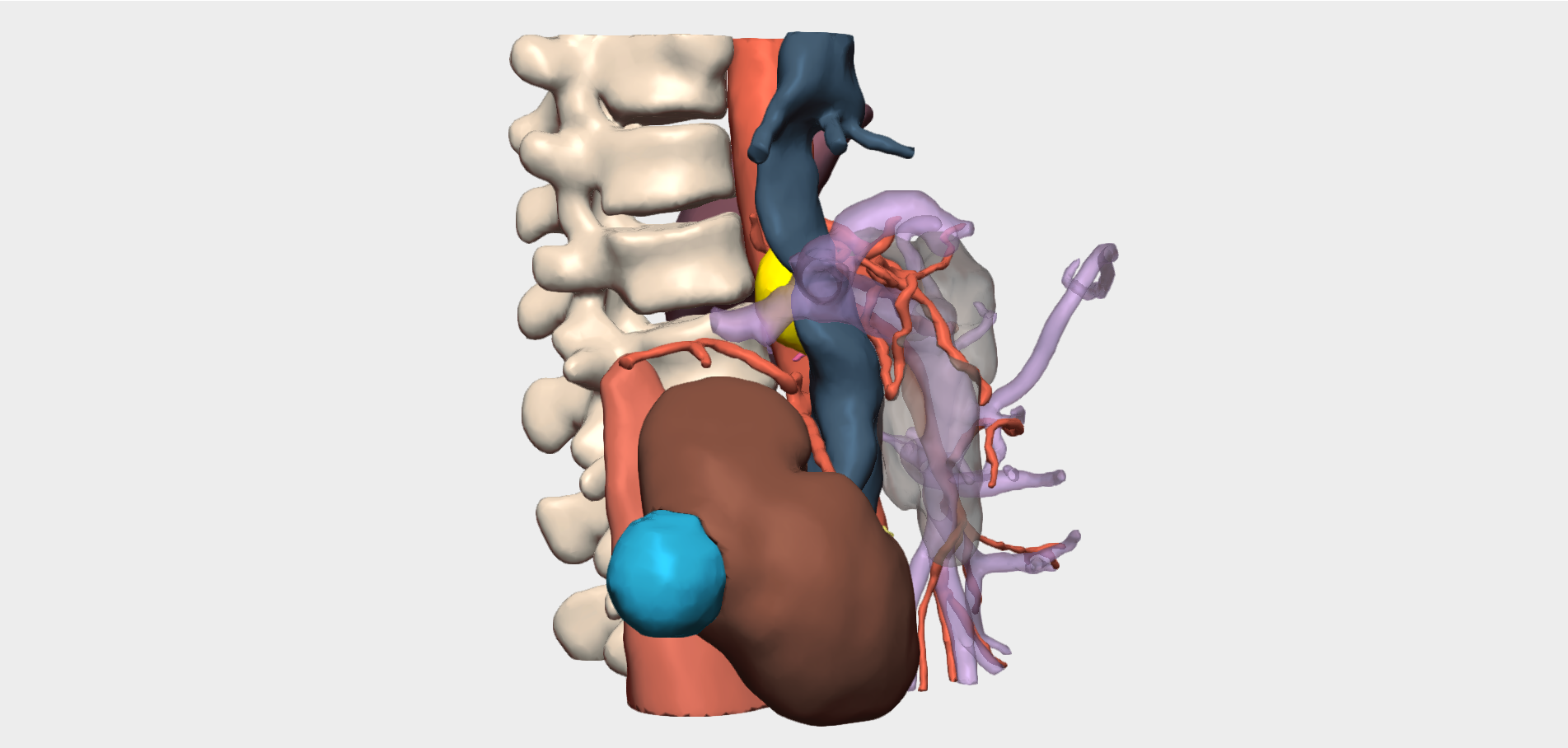
At a critical moment during the procedure, while the surgical team believed they were dissecting the healthy ureter, the 3D model was consulted intraoperatively to verify its position. The model confirmed that the left ureter, displaced by the size of the tumour, was located extremely close to the right ureter.
Thanks to this three-dimensional visualisation, the team was able to confirm correct dissection and avoid injury to the healthy ureter, significantly reducing the risk of complications.
Surgical Conclusions
In this liposarcoma case, use of the 3D model enabled a level of surgical precision that would not have been achievable with medical imaging alone, ensuring better surgical planning, reduced risk, and improved patient outcomes and recovery.
”The 3D model was extremely useful in understanding the lesion’s position, the structures involved, and the optimal surgical approach.
Dra. Yolanda QuijanoCo-Director of the Department of General and Digestive Surgery, Hospital Universitario HM Sanchinarro