Next Generation
3D modelling
SUCCESS STORY
Dr. Yolanda Quijano
Hospital Universitario HM Sanchinarro
Clinical Case
3D Reconstruction of a Pancreatic Tumour | Cella Medical Solutions
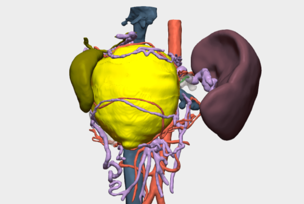
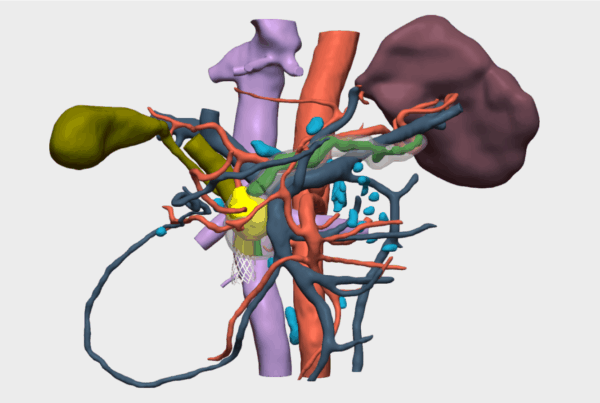
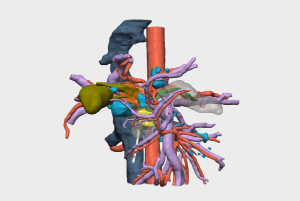
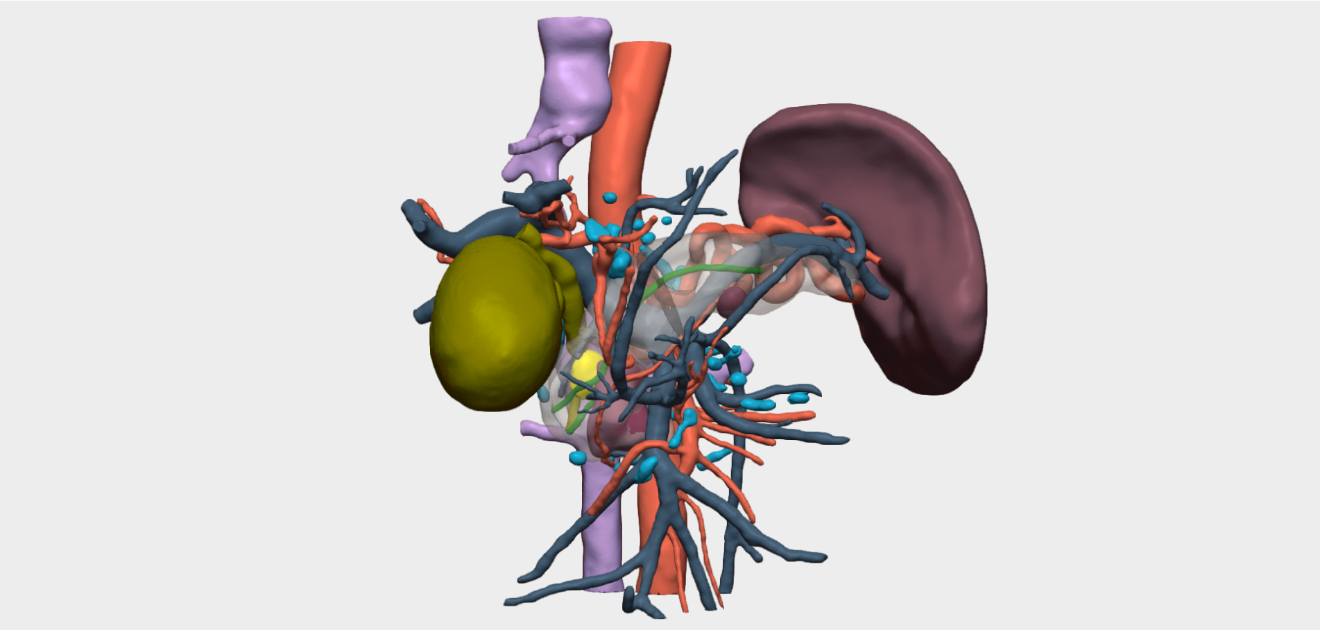
This case involves a 63-year-old male patient admitted with obstructive jaundice. Following comprehensive imaging studies, the hospital’s medical team identified a multiloculated cystic lesion located in the head of the pancreas, measuring approximately 42 × 35 × 20 mm.
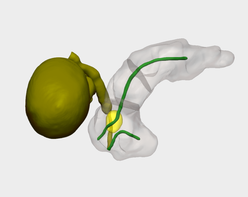
The lesion’s location significantly increased surgical complexity due to its close proximity to critical structures, including the pancreatic duct and the common bile duct (CBD). Careful preoperative planning was therefore essential to minimise the risk of complications and ensure optimal patient recovery.
3D Surgical Planning
During preoperative assessment, the surgical team needed to determine whether the biliary nodularity originated from the wall of the common bile duct or from the pancreatic parenchyma. To clarify this key diagnostic question, a patient-specific 3D model of the pancreatic anatomy was requested.
The 3D reconstruction revealed that the lesion originated in the common bile duct, with secondary involvement of the pancreas due to its location in the pancreatic head. This finding was critical in defining the surgical strategy and anticipating intraoperative findings.
Surgical Outcome
During surgery, Dr Yolanda Quijano encountered exactly what had been anticipated through the 3D model. Preoperative visualisation enabled precise identification of the lesion’s location and a clear understanding of its relationship with the common bile duct.
As a result, complete tumour resection was successfully achieved, leading to a favourable surgical outcome while minimising intraoperative risk for the patient.
”The 3D model clearly matched what we observed in the surgical field
Dr Yolanda QuijanoCo-Director of the Department of General and Digestive Surgery, Hospital Universitario HM Sanchinarro