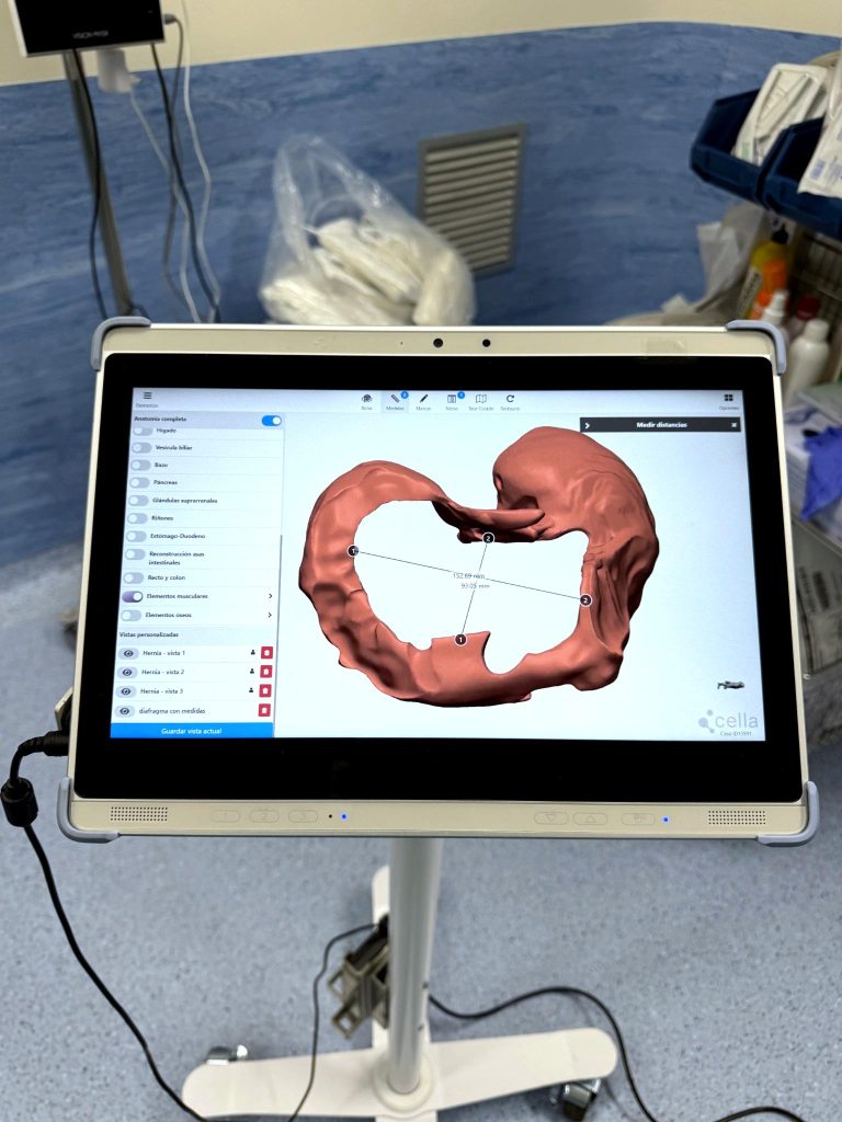
“3D reconstruction enabled a more precise understanding of the spatial arrangement of the anatomy, which was essential for planning the surgical management of a large diaphragmatic hernia”
Dr. Ángel Cuadrado – Consultant Specialist in General and Digestive Surgery, Hospital Universitario Infanta Sofía (Madrid)
Clinical Case
A 75-year-old female patient, former smoker, with a history of severe polytrauma in 2021 is presented.
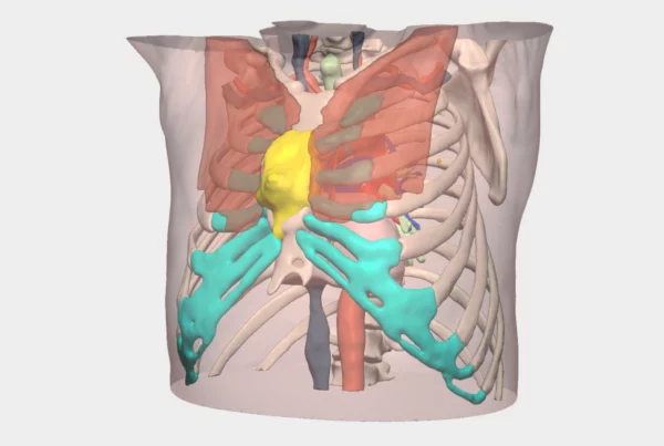
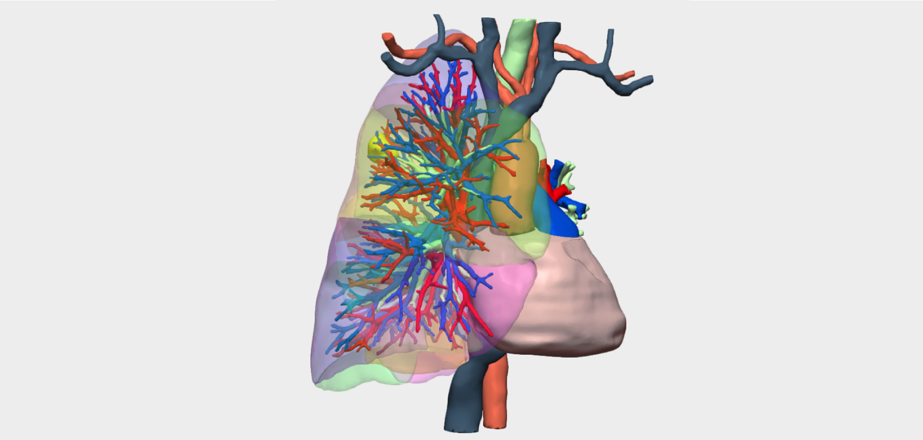
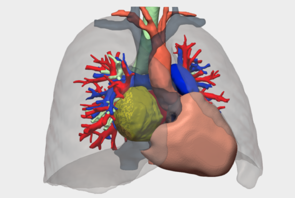
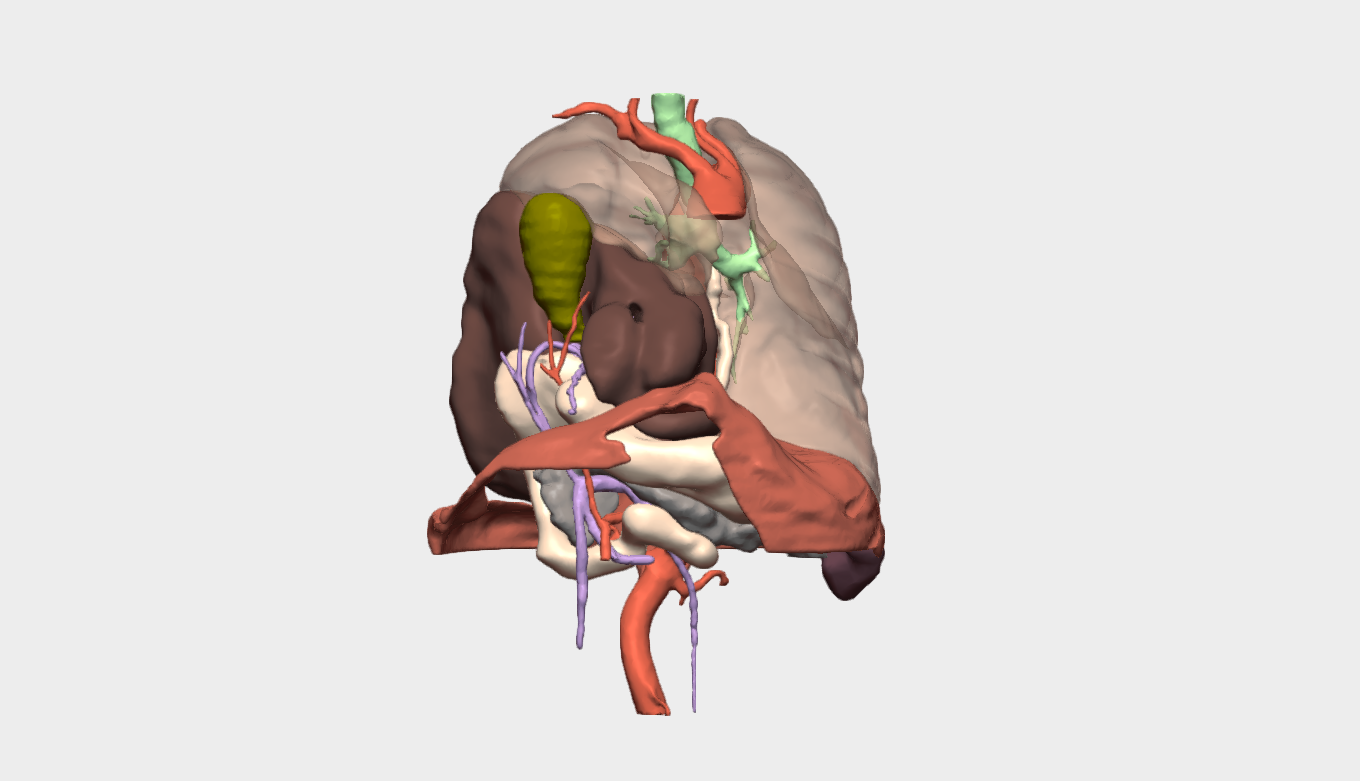
The patient had a large right diaphragmatic hernia with intrathoracic herniation of multiple abdominal structures, including the liver, gallbladder, colon, and gastric antrum. This visceral displacement caused a significant mass effect with mediastinal shift and right lower lobe atelectasis.
Additionally, cholelithiasis was identified, with suspicion of early acute cholecystitis.
The complexity of the case was mainly due to:
- Sequelae of prior polytrauma (rib fractures and ischiopubic ramus fractures), which could influence the surgical approach.
- The large size of the hernia.
- The extent of visceral content displaced into the thoracic cavity.
3D Planning
Given the description of a “large hernia” and the involvement of multiple organs (liver, pancreas, stomach), 3D anatomical reconstruction enabled an improved spatial understanding of the case.
Planning Tools
For the preoperative assessment, the following imaging studies were used:
- Abdominopelvic CT scan
- Thoracic MRI
Based on these imaging studies, a 3D model generated by Cella was also developed, supporting surgical planning. These assessments allowed accurate characterization of the herniated contents and evaluation of the mass effect on the mediastinum.
Surgical Strategy
After case evaluation, it was decided to perform, in a single surgical stage:
- Repair of the right diaphragmatic hernia.
- Cholecystectomy, to treat cholelithiasis with suspected early acute cholecystitis.
Surgical Outcomes
The immediate postoperative course was stable, although the patient developed a right pleural effusion that required thoracic drainage for 5 days. At discharge (December 9, 2025), the patient demonstrated good dietary tolerance and progressive clinical improvement.
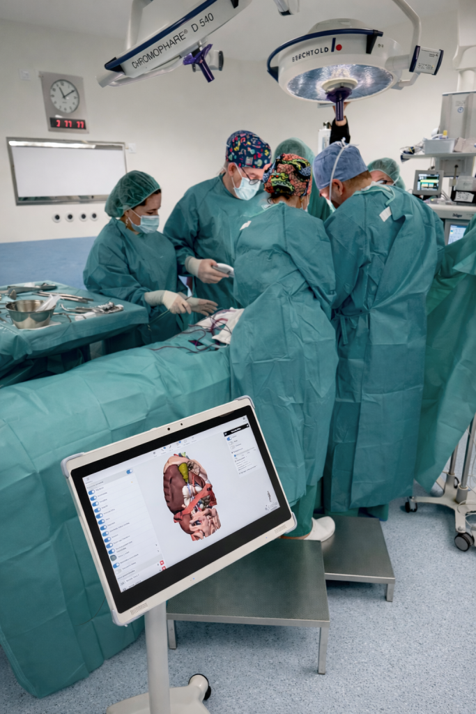
The surgery was successfully performed on December 1, 2025, by Dr. Ángel Cuadrado and Dr. Daniel Sánchez, from the General and Digestive Surgery team at Hospital Universitario Infanta Sofía (Madrid).
Follow us on social media to stay up to date with our latest updates!