
“Preoperative planning using 3D anatomical reconstruction enabled a more precise and conservative surgical strategy, facilitating a safe limited resection and avoiding an unnecessary major hepatectomy”.
Dr Pablo Beltrán Miranda – Clinical Lead, Specialist in General and Digestive Surgery at Hospital Juan Ramón Jiménez, Huelva.

Dr Teresa Moreno– Specialist in General and Digestive Surgery at Hospital Juan Ramón Jiménez, Huelva.
Dr Marcos Alba – Specialist in General and Digestive Surgery at Hospital Juan Ramón Jiménez, Huelva.
Clinical Case
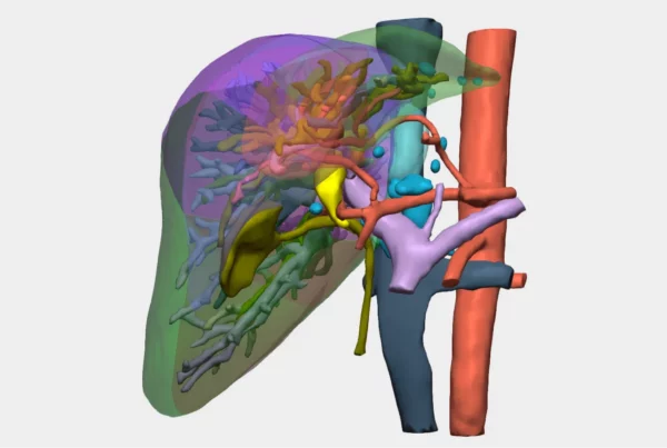
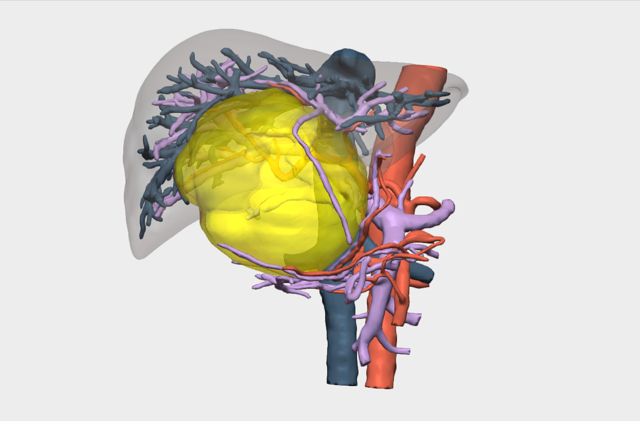
A 78-year-old patient was diagnosed with a multiloculated hepatic cystic lesion measuring approximately 10 × 10 cm, with high radiological suspicion of hepatic cystadenoma. The lesion involved liver segments IV, V, and VIII and caused compression of the portal bifurcation, with retrograde dilation of the intrahepatic biliary tree.
The main challenge of the case was the close relationship of the lesion with central vascular and biliary structures, which required careful surgical planning to minimize the risk of vascular or biliary injury and to avoid an unnecessary major hepatic resection.
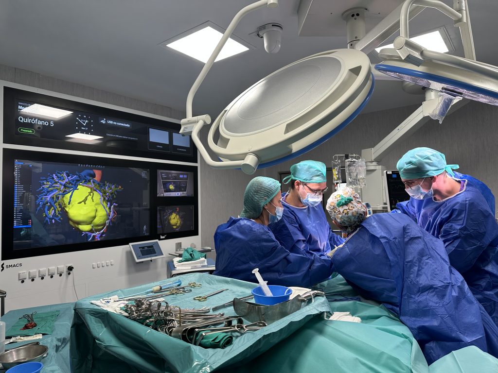
3D Planning
The 3D anatomical reconstruction allowed:
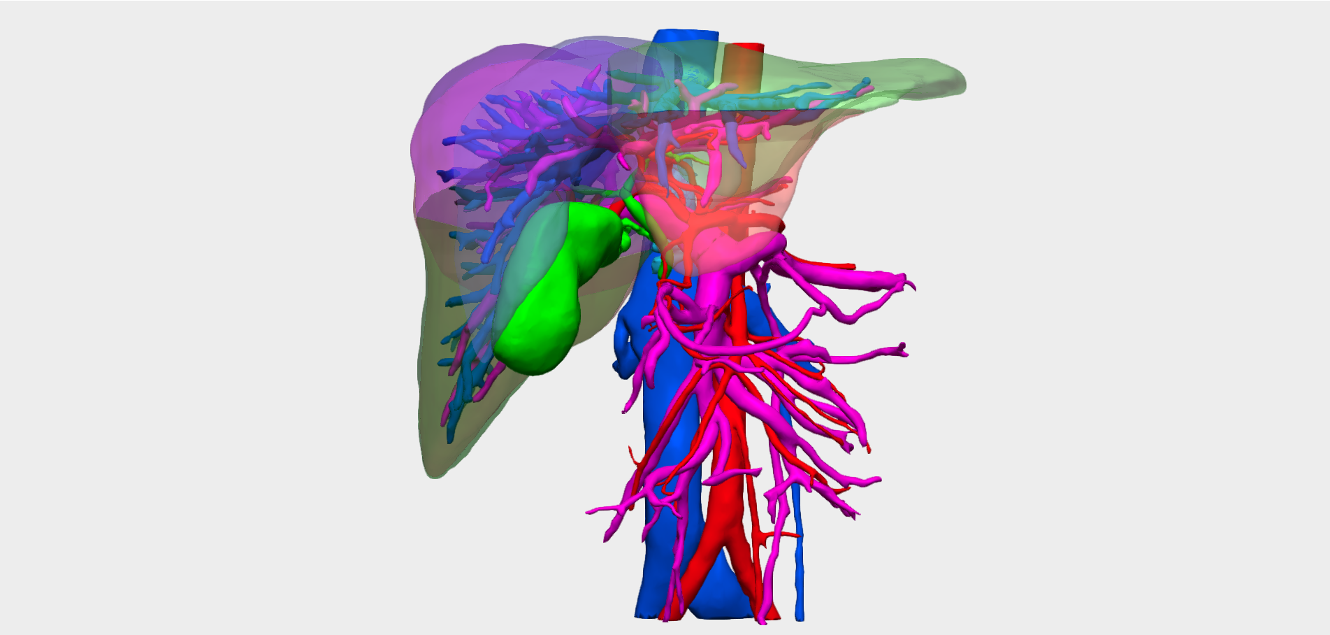
- Detailed analysis of the vascular and biliary relationships of the lesion.
- Identification of an arterial branch supplying segment IV in close contact with the tumor.
- Identification of an accessory bile duct draining into segment V.
This enabled precise spatial understanding of these structures and their relationship with the lesion, a key factor in surgical decision-making.
Vascular and biliary reconstructions were essential to assess the technical feasibility of enucleation.
Additionally, the ability to perform hepatic volumetry allowed estimation of the future liver remnant in case a major hepatectomy was required, which was particularly relevant given the patient’s age and the need to preserve adequate postoperative liver function.
Adopted Surgical Strategy
The initial strategy proposed was enucleation of the lesion, as the 3D model demonstrated contact, but no infiltration, of the main vascular structures.
However, central hepatectomy or extended right hepatectomy were considered as alternative surgical options in case vascular infiltration or technical impossibility of conservative resection was identified intraoperatively.
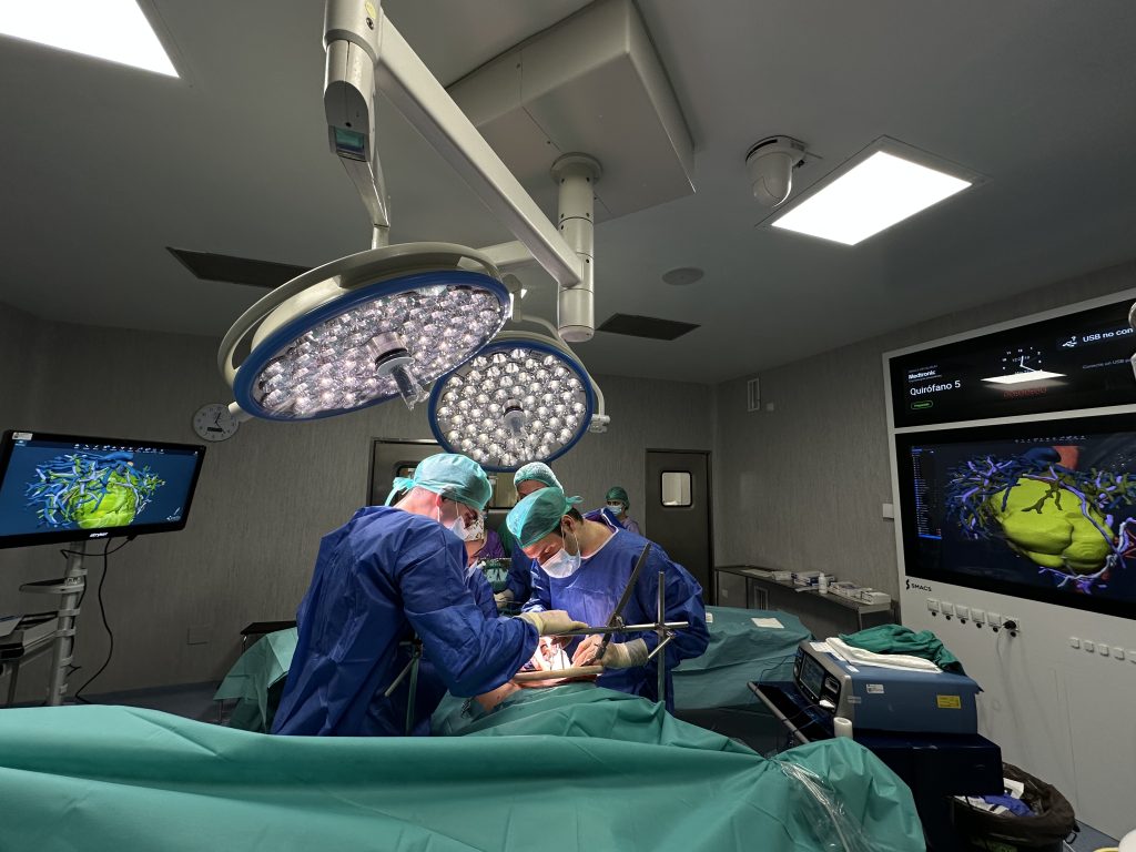
Surgical Outcomes
The procedure was carried out according to the planned strategy, and the following was performed:
- Complete enucleation of the lesion with preservation of the main vascular structures.
- Intraoperative identification and preservation of vascular and biliary branches in contact with the tumor.
The 3D model was key to identifying and preserving an accessory bile duct (segment VI), thereby preventing postoperative biliary complications.
Preoperative planning using 3D anatomical reconstruction enabled a more precise and conservative surgical strategy, facilitating a safe resection.
The surgery was performed by Dr. Pablo Beltrán, Head of the Hepatobiliary Surgery Section at Hospital Juan Ramón Jiménez in Huelva, together with Dr. María Teresa Moreno and Dr. Marcos Alba, attending surgeons of the HPB team.
Follow us on social media to stay up to date with our latest updates!