
“In this case, the Cella 3D model allowed us to plan the surgical approach to the large tumor mass by assessing its vascular supply and its relationship with critical structures”

Dr. Daniel Díaz Gómez, Consultant in General and Digestive Surgery, Virgen del Rocío Regional University Hospital (HUVR)
Case Presentation
We present the case of a 48-year-old female patient with no significant past medical history who developed persistent abdominal pain in the left upper quadrant, worsening while seated. She also reported menometrorrhagia.
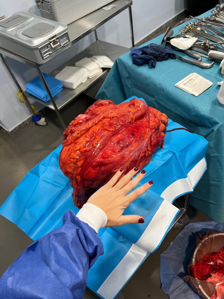
The patient was initially evaluated by the Gynecology Department, with no pathological findings identified. Subsequent abdominal ultrasound and CT urography revealed a giant abdominal mass measuring 24 × 14 × 24 cm, likely of retroperitoneal origin, displacing the left kidney, pancreas, stomach, and left colon.
Following preliminary diagnosis, a core needle biopsy was performed. Histopathological analysis confirmed a well-differentiated liposarcoma with nuclear expression of MDM2 and negative staining for beta-catenin, CD34, and S100.
After multidisciplinary evaluation by the HUVR Abdominal Sarcoma Tumor Board, a left-sided compartmental resection was planned, including resection of the giant tumor mass, distal splenopancreatectomy, left nephrectomy, left hemicolectomy, and possible gastrectomy.
Given the massive size of the tumor and the associated surgical risk, a three-dimensional model was requested to optimize preoperative planning and surgical strategy.
3D Surgical Planning and Approach
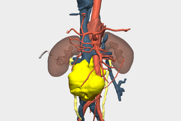
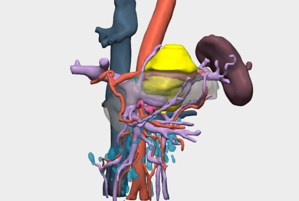
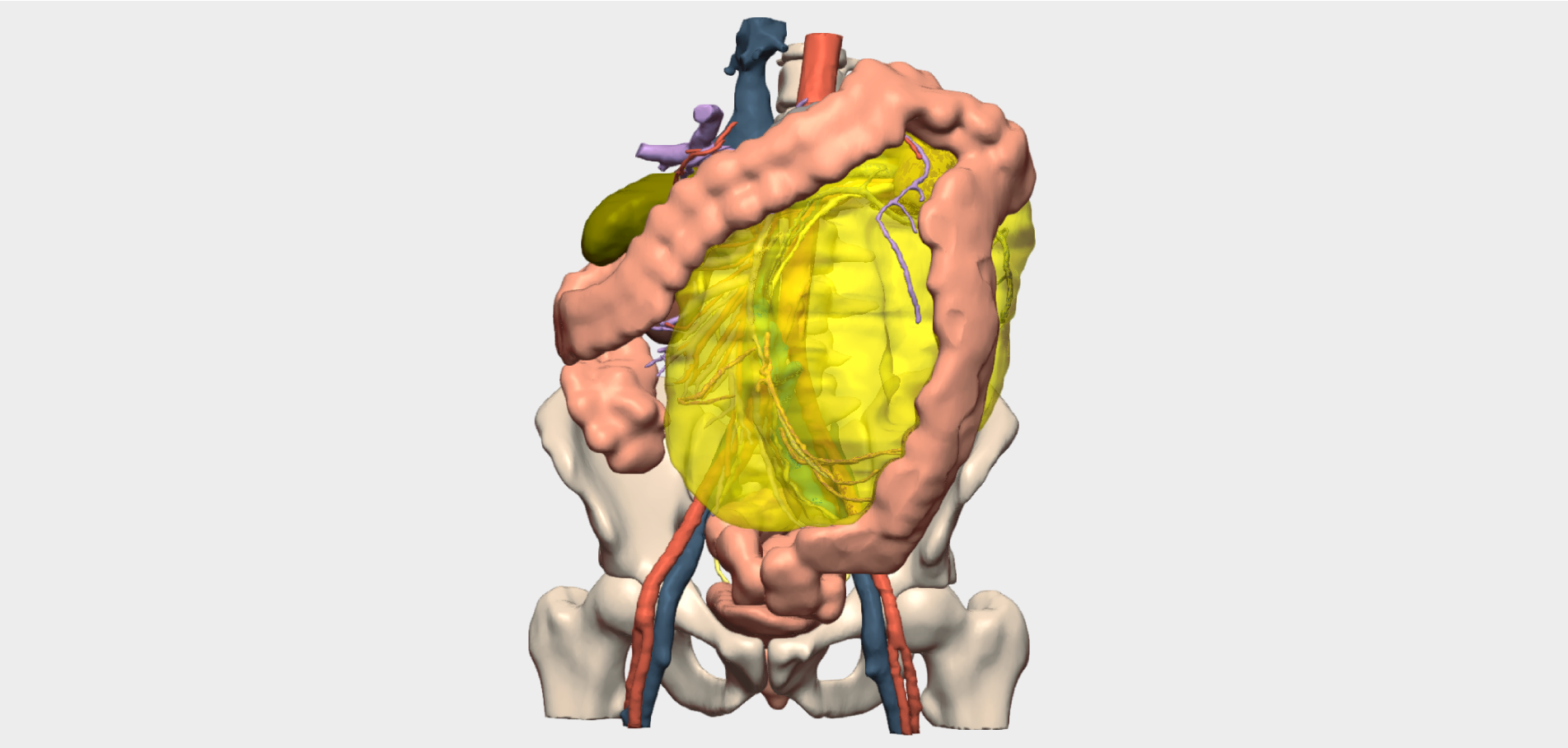
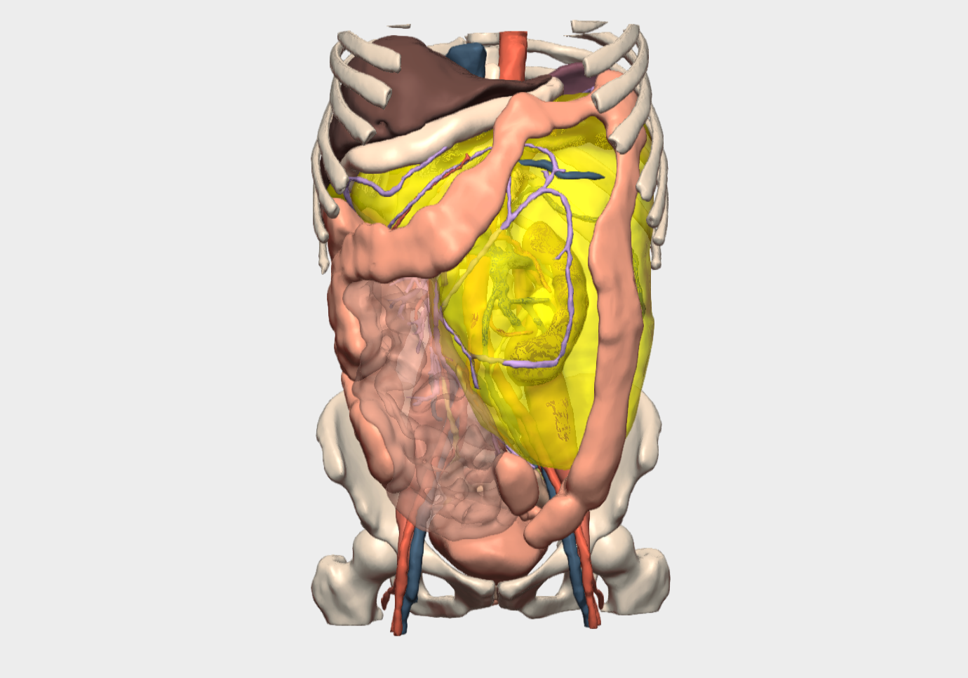
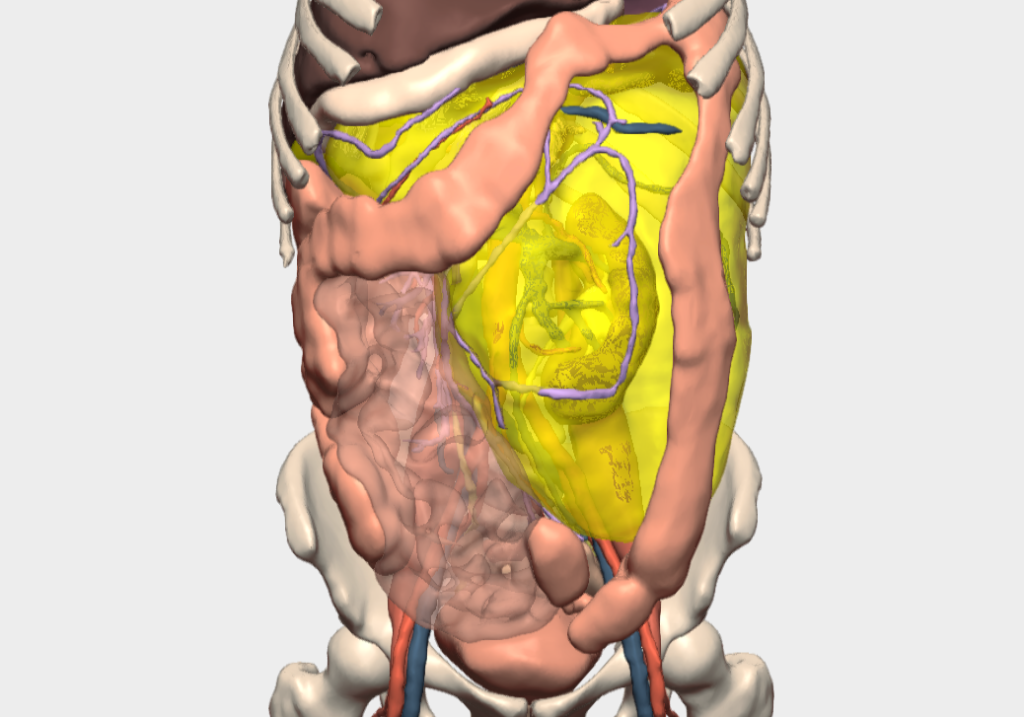
Considering the tumor’s anatomical characteristics and its relationships with adjacent organs, a 3D reconstruction was generated to allow for a more precise surgical approach. This technology enabled the surgical team to accurately visualize the mass in relation to vital structures such as the kidney, pancreas, and colon, thereby facilitating strategic operative planning.
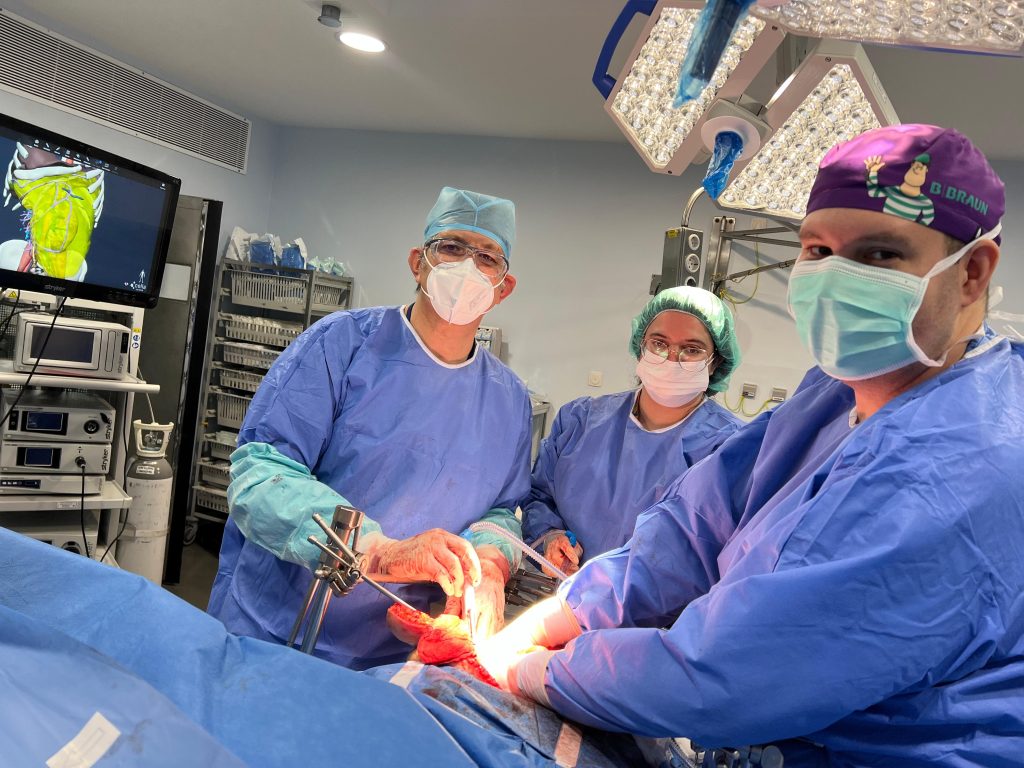
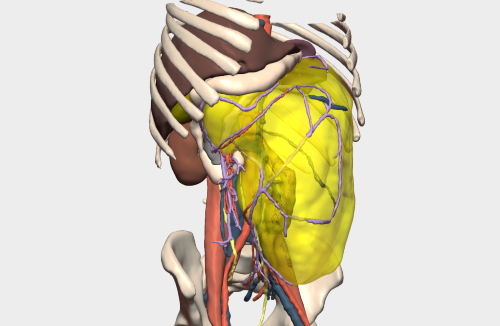
The 3D model was also used to evaluate the tumor’s vascular supply and its proximity to critical vascular structures. During surgery, particularly while approaching the renal hilum, the 3D model allowed precise identification of the left renal artery, which was located posterior to the left renal vein. This information proved crucial in enabling meticulous dissection, minimizing tissue manipulation, and enhancing operative safety.
Conclusions
This case of giant retroperitoneal liposarcoma highlights the advantages of advanced surgical planning in complex oncologic procedures. The use of a three-dimensional model contributed to reduced operative time, improved patient safety, and maintenance of high-quality surgical standards throughout the intervention.
¡Follow us on social media to stay up to date with our latest updates!