Clinical Case: Laparoscopic Heminephrectomy with 3D Surgical Planning
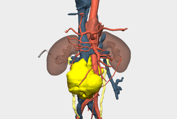
A 12-month-old female patient, weighing 9 kg, was followed by the Paediatric Oncology Service at the Complejo Hospitalario Universitario de Albacete due to a right renal duplication associated with a complex, multilobulated cystic tumour located in the upper renal moiety. The lesion was suggestive of a cystic nephroma and demonstrated progressive growth on follow-up ultrasound, reaching an approximate size of 3 × 3 × 2 cm at the time of surgical evaluation.
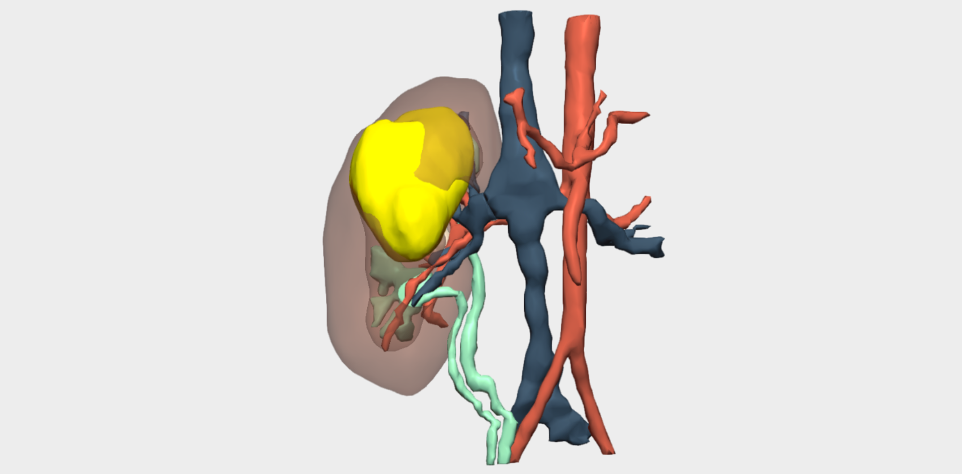
Following multidisciplinary assessment by the Oncology Committee, a right upper pole laparoscopic heminephrectomy was indicated for diagnostic histopathological analysis. Imaging studies were inconclusive and did not allow definitive diagnosis or exclusion of malignancy. Additional imaging performed at the Complejo Hospitalario Universitario de Albacete, including MRI and angio-CT, anticipated significant challenges in vascular localisation and dissection. The renal artery and vein bifurcated within the kidney, beneath the tumour and in close contact with it. Furthermore, the small calibre of these vessels limited precise visualisation of their anatomical course.
Given the rarity of the case and the anatomical complexity, the use of a patient-specific 3D model based on the most recent imaging studies was considered essential.
3D Surgical Planning: Model Utility and Key Functionalities
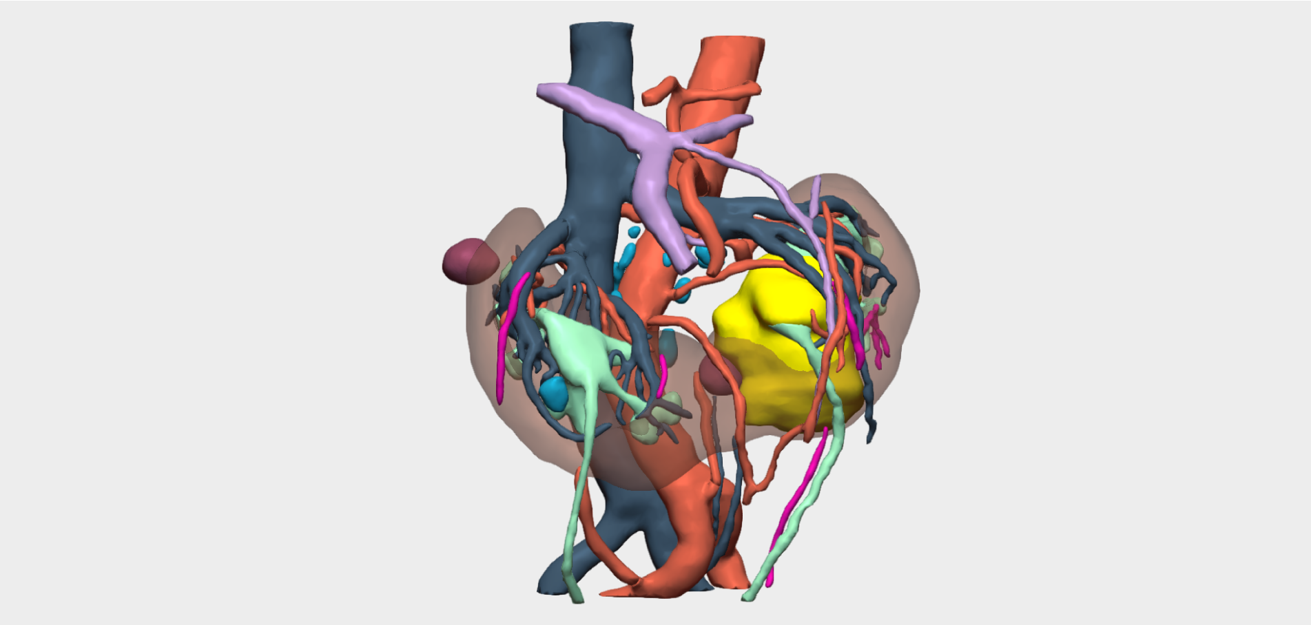
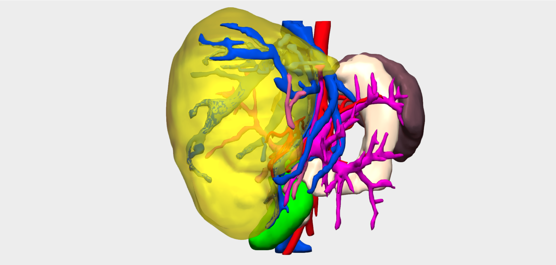
Three-dimensional reconstruction enabled precise localisation of the vascular bifurcation, revealing the presence of two arteries and two veins supplying each renal moiety —an anatomically relevant finding that had not been previously identified and was critical for surgical planning.


Detailed 3D anatomical analysis, combined with advanced functionalities such as arterial territory assessment and the ability to work intraoperatively with dual visualisation —3D reconstruction and laparoscopy displayed simultaneously— was a key factor in the success of the procedure.
Surgical Outcome and Conclusions
The surgery was completed successfully following selective vascular control, without clamping of the renal hilum, thereby minimising the risk of injury to the healthy renal moiety. No intraoperative complications were observed.
Postoperatively, the patient remained in the Paediatric Intensive Care Unit for 24 hours and was discharged on postoperative day six following removal of the surgical drain, which had been placed due to a minor urinary leak.
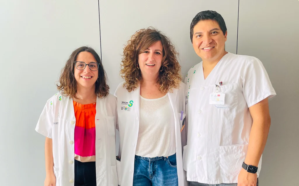
The right upper pole laparoscopic heminephrectomy was performed by the Paediatric Urology Unit of the Complejo Hospitalario Universitario de Albacete, with Dr Verónica Marijuán Sahuquillo as lead surgeon, assisted by Dr María Ramírez Piqueras and Dr Javier Rojas Ticona, under the supervision of the Head of Department, María Soledad Fernández.
To sum it up, in this case, 3D reconstruction enabled detailed visualisation of the renal vascular anatomy, facilitating preoperative planning and optimising the surgical strategy. This approach reduced intraoperative morbidity, shortened operative time and, most importantly, preserved the healthy renal moiety.
¡Follow us on social media to stay up to date with our latest updates!