
3D models for Hepatobiliary Surgery
General and Digestive System Surgery
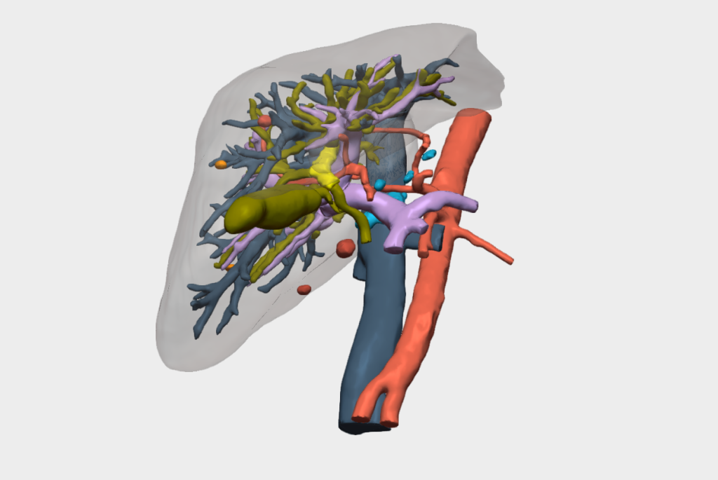
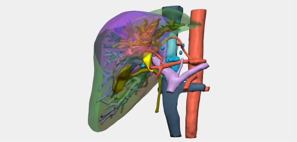
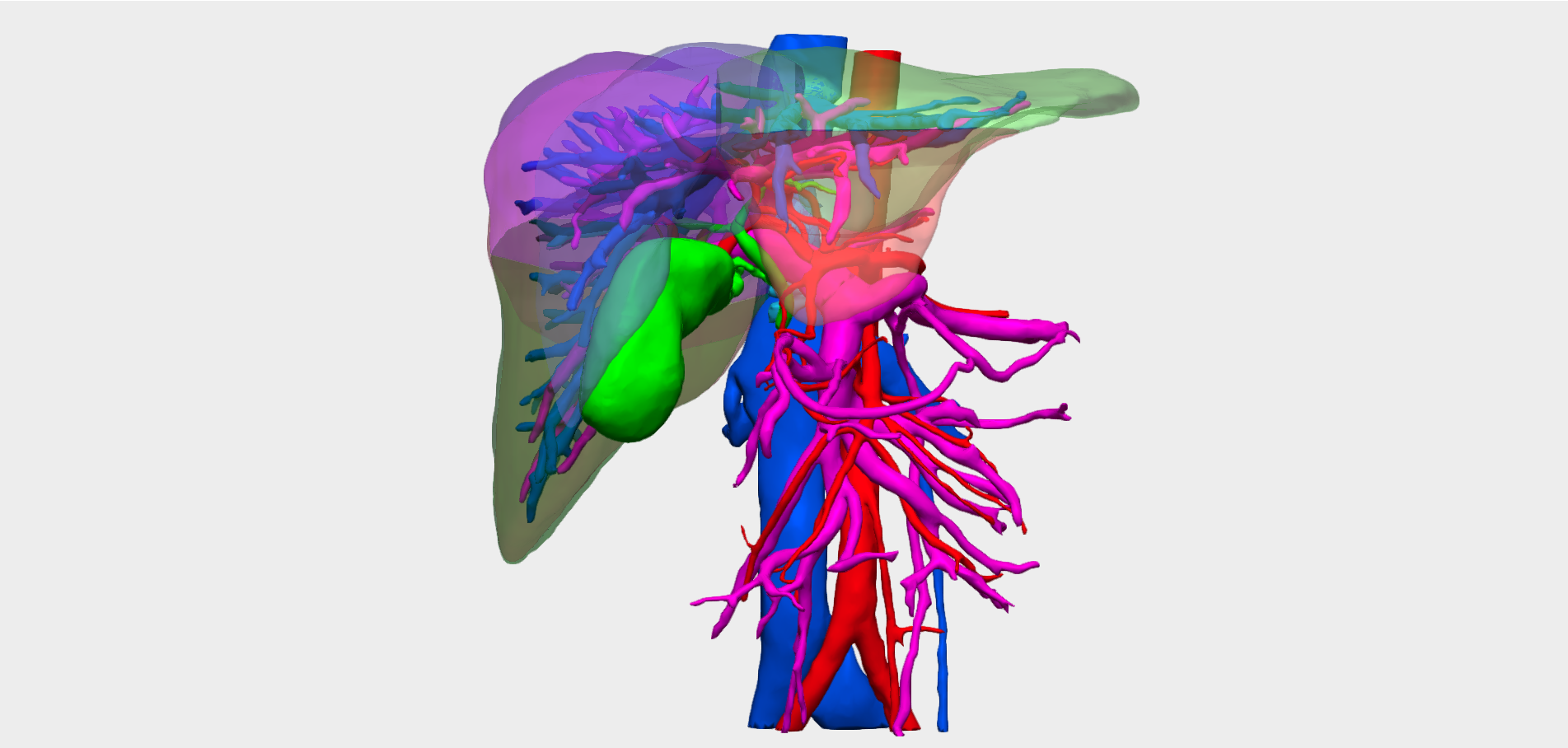
In Hepatobiliary Surgery, precision and planning are essential for performing highly complex procedures such as liver resections or tumour excisions. Our 3D models provide a comprehensive reconstruction of the hepatic, biliary and vascular structures, supporting preoperative planning and reducing surgical risks.


An advanced solution for planning complex liver and biliary surgeries, enhancing clinical outcomes and enabling new surgical possibilities.
- Higher proportion of negative resection margins
- Helps predict vascular invasion
- Stimulates the design of new surgical techniques
- Reduction in intraoperative bleeding
- Accurate calculation of liver and tumour volumes
- Better understanding of liver anatomy and spatial relationships
Specific tools for 3D planning of Hepatobiliary Surgery
We collaborate with hepatobiliary surgeons to develop dedicated tools
Our 3D models offer features tailored to real clinical needs, enabling personalised surgical planning for across specialties and pathologies.

Resection simulation
Plan subsegmentectomies, ruled and unruled resections, and tumourectomies.

Biliary variants
Shows a detailed analysis of biliary variants and the Bismuth-Corlette classification.

Volumetric study
Details of the total and remaining volume of segments and lesions.

Creation of vascular territories
Customised vascular territories to improve understanding of vascularisation.
And more…
Clinical indications in Hepatobiliary Surgery with 3D models
Especially recommended for conditions and procedures in which preoperative planning can optimise outcomes and enhance surgical safety.
LIVER
Primary Malignant Tumours
- Hepatocellular carcinoma (HCC)
- Intrahepatic cholangiocarcinoma
- Hepatic angiosarcoma
Malignant Metastatic Tumours
- Colorectal metastases
- Breast, lung, or pancreatic metastases
Benign or Premalignant Lesions
- Large liver adenomas (>5 cm)
- Focal nodular hyperplasia (FNH)
- Giant liver haemangiomas
Cystic and Congenital Liver Diseases
- Simple Complicated Liver Cysts
- Hydatid Cysts
- Polycystic Liver Disease
Vascular Pathologies
- Portal vein thrombosis
- Budd-Chiari syndrome
- Hepatic arterial aneurysms
BILE DUCT
Malignant Tumours
- Hilar cholangiocarcinoma (Klatskin)
- Choledochal carcinoma
- Tumours of the ampulla of Vater
Complex Gallstones
- Intrahepatic lithiasis
- Complicated cholelithiasis
Inflammatory and Benign Conditions
- Benign biliary stenosis
- Choledochal cysts
- Caroli’s disease
GALLBLADDER
Malignant Tumours
- Gallbladder Carcinoma
- Large gallbladder polyps (>10 mm)
Inflammatory or Complicated
- Complicated Acute Cholecystitis
- Mirizzi Syndrome
COMBINED OR SPECIAL
Liver Transplant
- Liver transplant from living donor
- Cadaveric donors with complex anatomies
Liver and Biliary Trauma
- Complex Trauma Injuries
Mixed and Rare Tumours
- Hepatoblastoma in Adults
- Biphasic or mixed tumours
Infections
- Extensive Liver Abscesses
Testimonials from surgeons who already use our 3D models
”With the 3D model, we can accurately determine the dimensions of the tumour, detecting any involvement of other organs or vascular structures to avoid unexpected complications during the operation.
Dr. Manuel BarreraHead of General and Digestive Surgery. Hospiten Rambla University Hospital, Tenerife.
”The model allowed us to confirm the anatomy, assess the relationship between the tumour and the aberrant artery, and verify the absence of vascular invasion.
Dr. Miguel Ángel Gómez BravoHPB and Transplant Surgeon. Virgen del Rocío University Hospital, Seville.


Clinical cases in hepatobiliary cancer surgery
Learn about real success stories
Request them additionally
3D Printing models
They act as a haptic extension in the operating theatre. Each one undergoes a rigorous quality control and radiological validation process to ensure its accuracy.
- Real scale
- Haptic interpretation of anatomy
- Structure differentiation by colours
- Advanced modelling
- Sterilisable using standard methods (H₂O₂)
- Ready for rapid delivery (<72 hours)
- Intended for research and teaching purposes only

© 2026 Cella Medical Solutions