
3D models for Thoracic Surgery
Thoracic Surgery
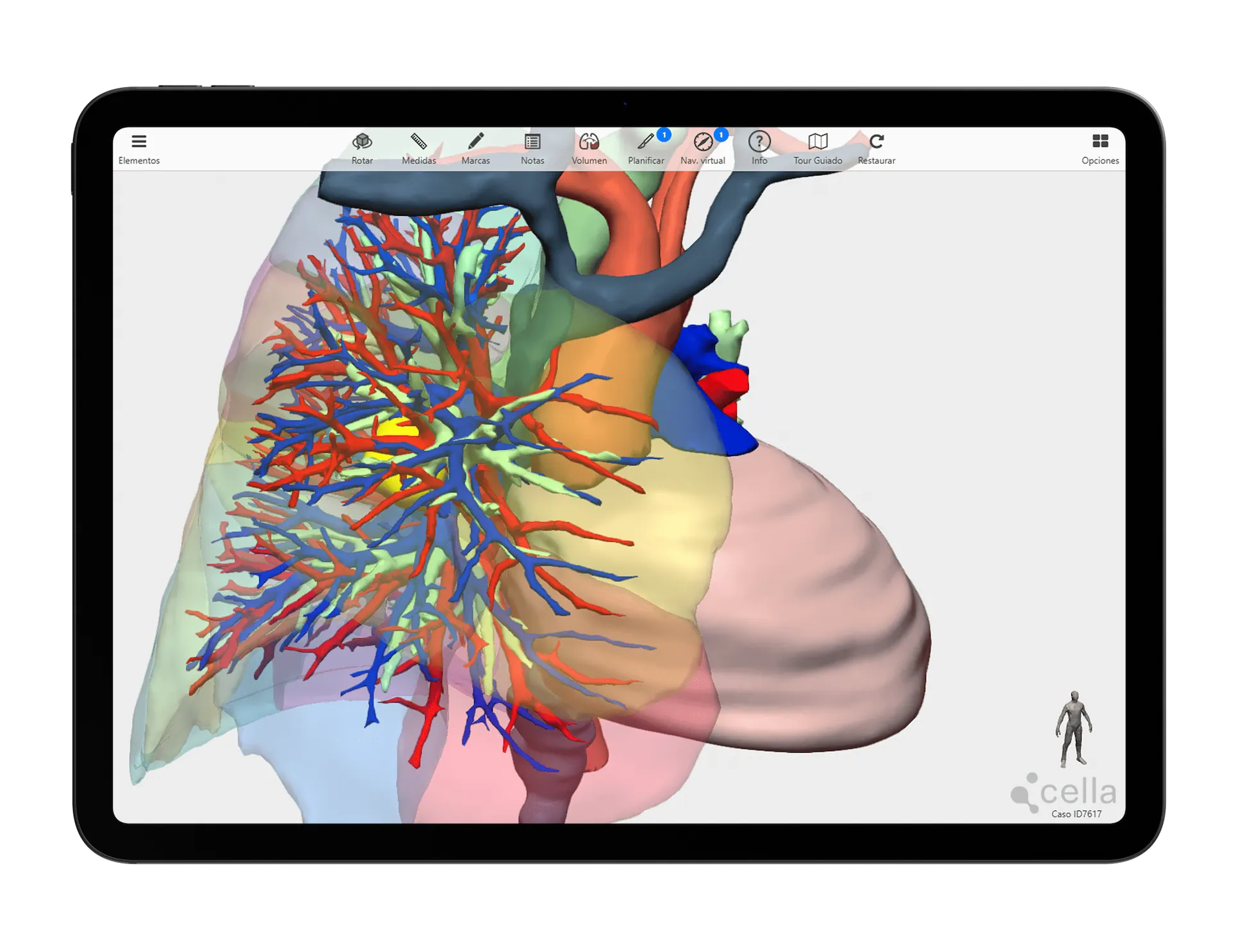
Thoracic Surgery involves treating pathologies in a region containing vital organs such as the lungs, heart, oesophagus and major vessels, enclosed by the chest wall and its musculature. 3D models enable precise localisation of lesions and their relationship to critical structures.
An advanced solution for planning complex thoracic surgeries, enhancing clinical outcomes and enabling new surgical possibilities.
- Recommended for use in segmentectomies
- Reduces the number of stapler reloads and the air leak rate
- Facilitates vascular assessment and lesion localisation
- Enables minimally invasive resections to be performed with greater precision and safety
- Allows assessment of resectability, resection margins and identification of anatomical variations
Specific tools for 3D planning of Thoracic Surgery
We collaborate with thoracic surgeons to develop dedicated tools.
Our 3D models offer features tailored to real clinical needs, enabling personalised surgical planning for across specialties and pathologies.
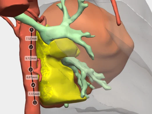
Measure distances
Measure the distance between points of interest or the diameter of elements such as tumours or vascularisation.
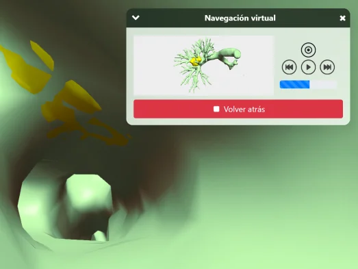
Intraductal and intravascular virtual navigation
Visualise the interior of the airway or vascularisation to analyse tumour invasion.
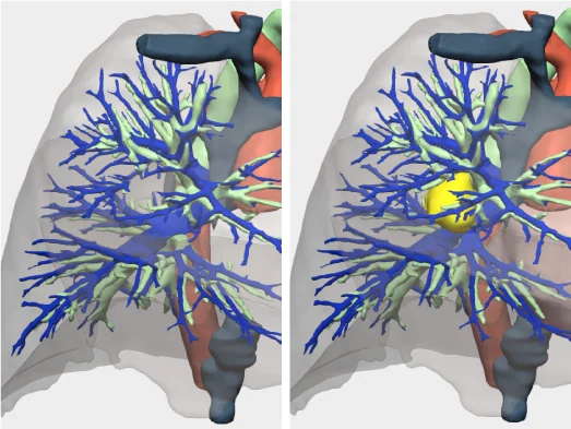
Resection planning
Simulate tumour resections by territory, lobectomies, tumourectomies, or subsegmentectomies.
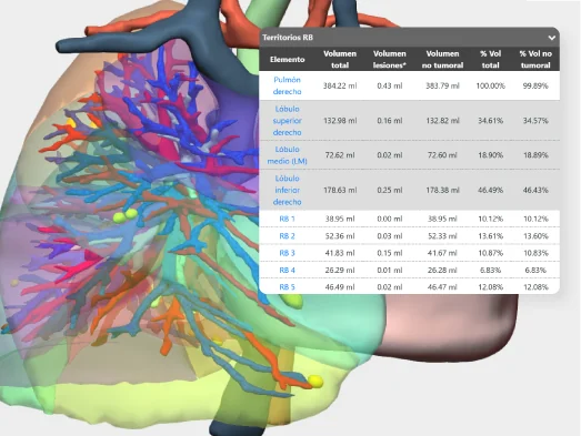
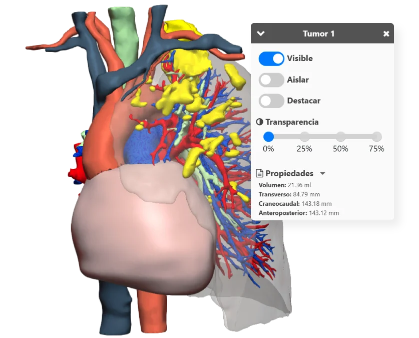
Volumetric study
Details of the total and remaining volume of segments and lesions.
And more…
Clinical indications in Thoracic Surgery with 3D models
Especially recommended for conditions and procedures in which preoperative planning can optimise outcomes and enhance surgical safety.
LUNG AND AIRWAY
Lung tumours
- Primary lung cancer (non-small cell carcinoma and small cell carcinoma).
- Lung metastases from tumours in other organs.
- Benign tumours (hamartomas, bronchial adenomas, etc.).
Infectious/inflammatory diseases
- Complicated pneumonia (lung abscesses).
- Bronchiectasis (chronic dilation of the bronchi).
- Tuberculosis and cavitary sequelae.
Obstructive and congenital pathologies
- Chronic Obstructive Pulmonary Disease (COPD) and emphysematous bullae
- Congenital malformations (pulmonary sequestration, bronchogenic cyst, etc.)
- Pulmonary trauma
MEDIASTINUM
Mediastinal masses
- Thymic tumours (thymoma, thymic carcinoma)
- Lymphoma (Hodgkin’s and non-Hodgkin’s) with mediastinal involvement
- Mediastinal cysts (bronchogenic, congenital pericardial, oesophageal)
- Intrathoracic thyroid (substernal goitre)
Pathologies of the oesophagus (intrathoracic portion)
- Oesophageal tumours (adenocarcinoma, squamous cell carcinoma) and complications (fistulas, stenosis)
- Oesophageal diverticula (cervical Zenker’s, epiphrenic)
- Motor disorders (achalasia, diffuse spasm) with repercussions in the mediastinum
Mediastinal lymphadenopathy
- Metastatic (from lung tumours or other tumours)
- Inflammatory (sarcoidosis, lymph node tuberculosis)
Others
- Mediastinal trauma
CHEST WALL AND DIAPHRAGM
Chest wall tumours
- Primary tumours (soft tissue sarcomas, rib chondrosarcomas, osteosarcomas).
- Metastases in ribs or sternum.
Chest wall deformities
- Pectus excavatum and pectus carinatum
- Poland Syndrome (partial agenesis of the pectoral muscle)
Chest wall trauma
- Multiple rib fractures, sternal fractures and their complications (unstable chest)
- Post-traumatic or post-surgical reconstructions (tumour resection)
Diaphragmatic hernias
- Congenital (Bochdalek, Morgagni)
- Acquired (traumatic)
Testimonials from surgeons who already use our 3D models
”It allowed us to identify the patient's vascular variants and the precise boundaries of segment 2, ensuring an adequate resection margin.
Dr. Santiago FigueroaThoracic Surgeon. Valencia Clinical Hospital


Clinical cases in Thoracic Cancer Surgery
Learn about real success stories
Request them additionally
3D Printing models
They act as a haptic extension in the operating theatre. Each one undergoes a rigorous quality control and radiological validation process to ensure its accuracy.
- Real scale
- Haptic interpretation of anatomy
- Structure differentiation by colours
- Advanced modelling
- Sterilisable using standard methods (H₂O₂)
- Ready for rapid delivery (<72 hours)
- Intended for research and teaching purposes only

© 2026 Cella Medical Solutions