
3D models for Peritoneal and Retroperitoneal Surgery
General and Digestive System Surgery
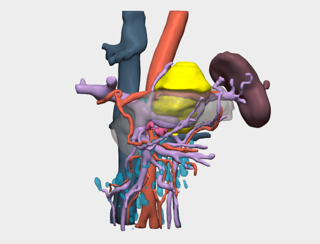
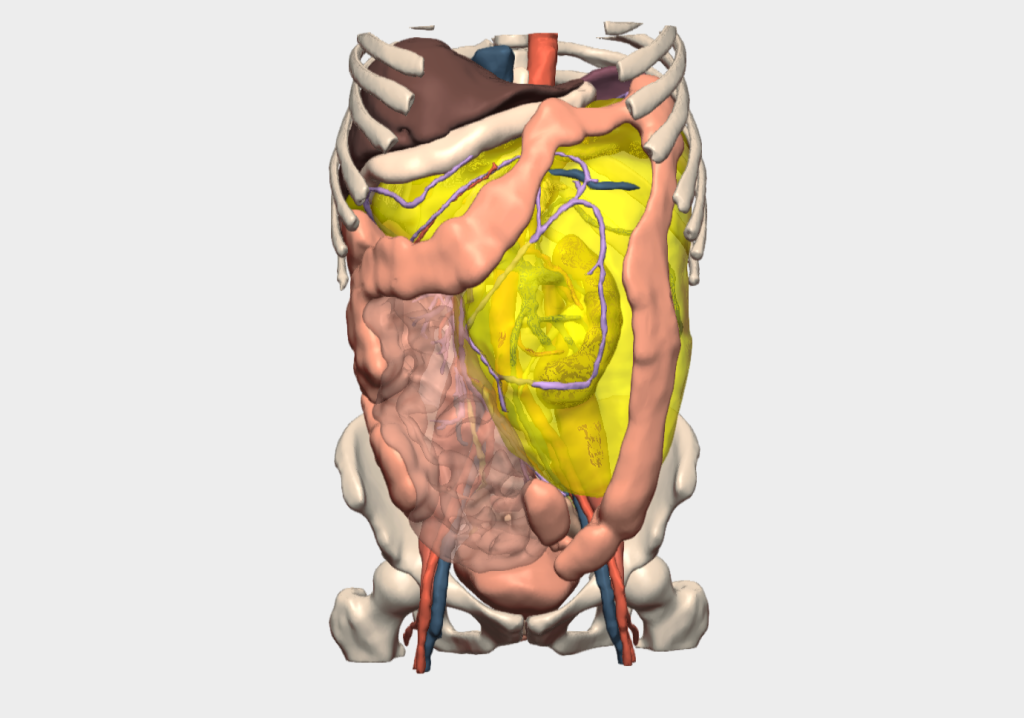
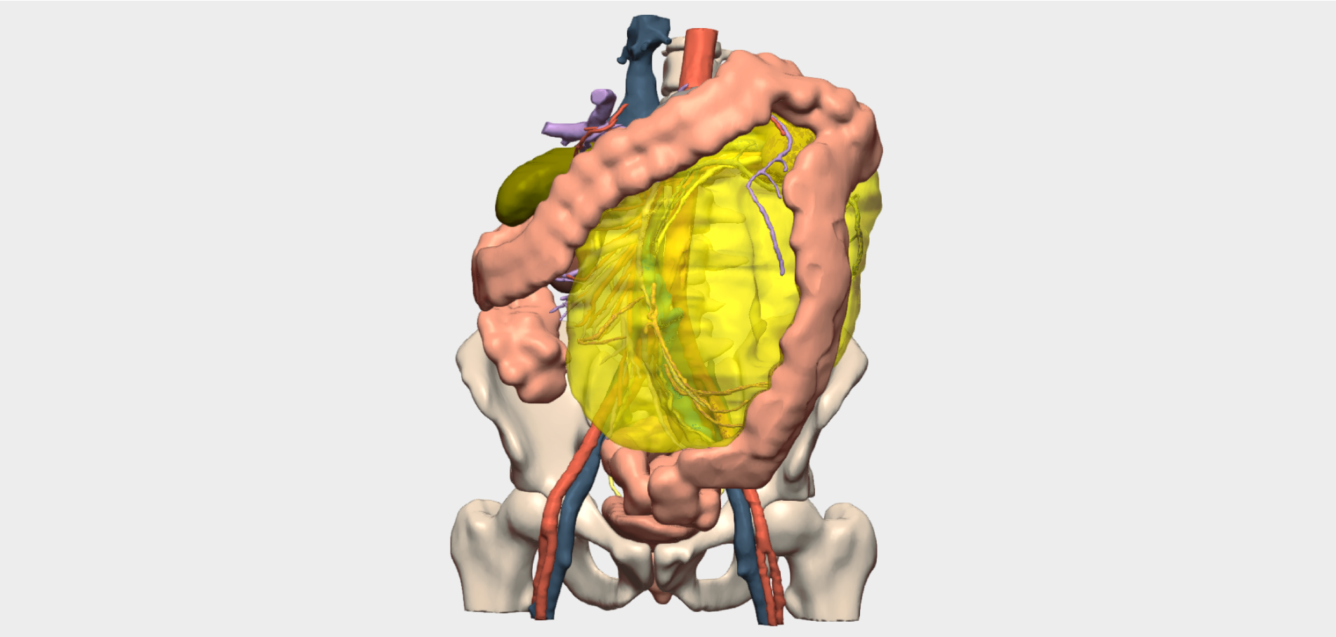
Peritoneal and Retroperitoneal Surgery is often characterised by the large size of lesions, which can involve vital adjacent organs and structures. 3D models enable surgeons to assess tumour extent, analyse anatomical relationships and plan the optimal surgical approach.


An advanced solution for planning complex peritoneal and retroperitoneal surgeries, enhancing clinical outcomes and enabling new surgical possibilities.
- Better understanding of the patient’s anatomy and less cognitive effort required by the surgeon to analyse structures
- Useful tool in the education and training of residents
- Less intraoperative blood loss
- Facilitates interdisciplinary communication between doctors
- Reduction in intraoperative complications
Specific tools for 3D planning in Peritoneal and Retroperitoneal Surgery
We collaborate with leading surgeons to develop functionalities
Our 3D models offer features tailored to real clinical needs, enabling personalized surgical planning for each specialty and pathology.

Relationship between segments and carcinomatosis
Shows the patient’s abdominal regions and the relationship of the lesion to each of them.

Relationships between pathological elements
Shows possible infiltrations/contacts between the tumour and other elements.

Minimum distances
Shows minimum distances between tumour elements and other elements.

Measure distances
Measure the distance between points of interest or the diameter of elements such as tumours or vascularisation.
And more…
Clinical indications in Peritoneal and Retroperitoneal Surgery with 3D models
Especially recommended for conditions and procedures in which preoperative planning can optimise outcomes and enhance surgical safety.
Peritoneal pathologies
- Peritonitis
- Peritoneal carcinomatosis
- Ascites
- Peritoneal mesothelioma
- Peritoneal cysts and pseudocysts
- Peritoneal endometriosis
- Peritoneal abscesses
- Abdominal wall hernias
Retroperitoneal pathologies
- Primary retroperitoneal tumours
- Lipomas and liposarcomas
- Leiomyomas and leiomyosarcomas:
- Mesenchymal tumours
- Retroperitoneal lymphoma
- Retroperitoneal fibrosis (Ormond’s disease)
- Retroperitoneal haematoma
- Retroperitoneal abscess
- Retroperitoneal pancreatic pathologies
Testimonials from surgeons who already use our 3D models
”With the 3D model, we can accurately determine the dimensions of the tumour, detecting any involvement of other organs or vascular structures to avoid unexpected complications during the operation.
Dr. Manuel BarreraHead of General and Digestive Surgery. Hospiten Rambla University Hospital, Tenerife.
”In sarcoma surgery, where oncologic margins and the relationship with critical structures are decisive, Cella’s 3D models provide invaluable support. They allow precise understanding of tumor extent, its relationship with vessels, nerves, and adjacent organs, and enable a much safer and more personalized resection strategy. Through this advanced planning, surgical margins can be optimized, operative time reduced, and complications minimized—facilitating less aggressive procedures and better functional preservation for the patient.
Dr. DíazHead of General and Digestive Surgery. Hospiten Rambla University Hospital, Tenerife.
Clinical cases in Peritoneal and Retroperitoneal Cancer Surgery
Learn about real success stories
Request them additionally
3D Printing models
They act as a haptic extension in the operating theatre. Each one undergoes a rigorous quality control and radiological validation process to ensure its accuracy.
- Real scale
- Haptic interpretation of anatomy
- Structure differentiation by colours
- Advanced modelling
- Sterilisable using standard methods (H₂O₂)
- Ready for rapid delivery (<72 hours)
- Intended for research and teaching purposes only

© 2026 Cella Medical Solutions