
3D models for Paediatric Surgery
Cella Surgical Planner
Paediatric procedures present challenges due to the small size and anatomical complexity of the patients. 3D models for paediatric surgery enable in-depth anatomical knowledge, the identification of variants, and communication with the family and patient.

Our product portfolio
Oncological surgery
Urological surgery
Digestive surgery
Hepatobiliary surgery
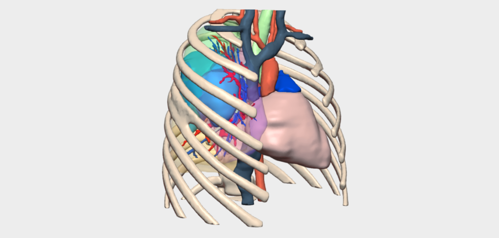
Thoracic surgery
Plastic surgery
Perinatal surgery

An advanced solution for planning complex paediatric surgeries, enhancing clinical outcomes and enabling new surgical possibilities.
- Reduction in fluoroscopic exposure and radiation
- Improved doctor-patient/family communication
- Improved surgical management of complex tumours and safer resections
- Enables conservative nephron-sparing surgery, avoiding nephrectomies
- Helps optimise the surgical plan by influencing therapeutic decisions
- Provides insight into anatomical variations
Specific tools for 3D planning of Paediatric Surgery
We collaborate with paediatrics surgeons to develop dedicated tools
Our 3D models offer features tailored to real clinical needs, enabling personalized surgical planning for each specialty and pathology.

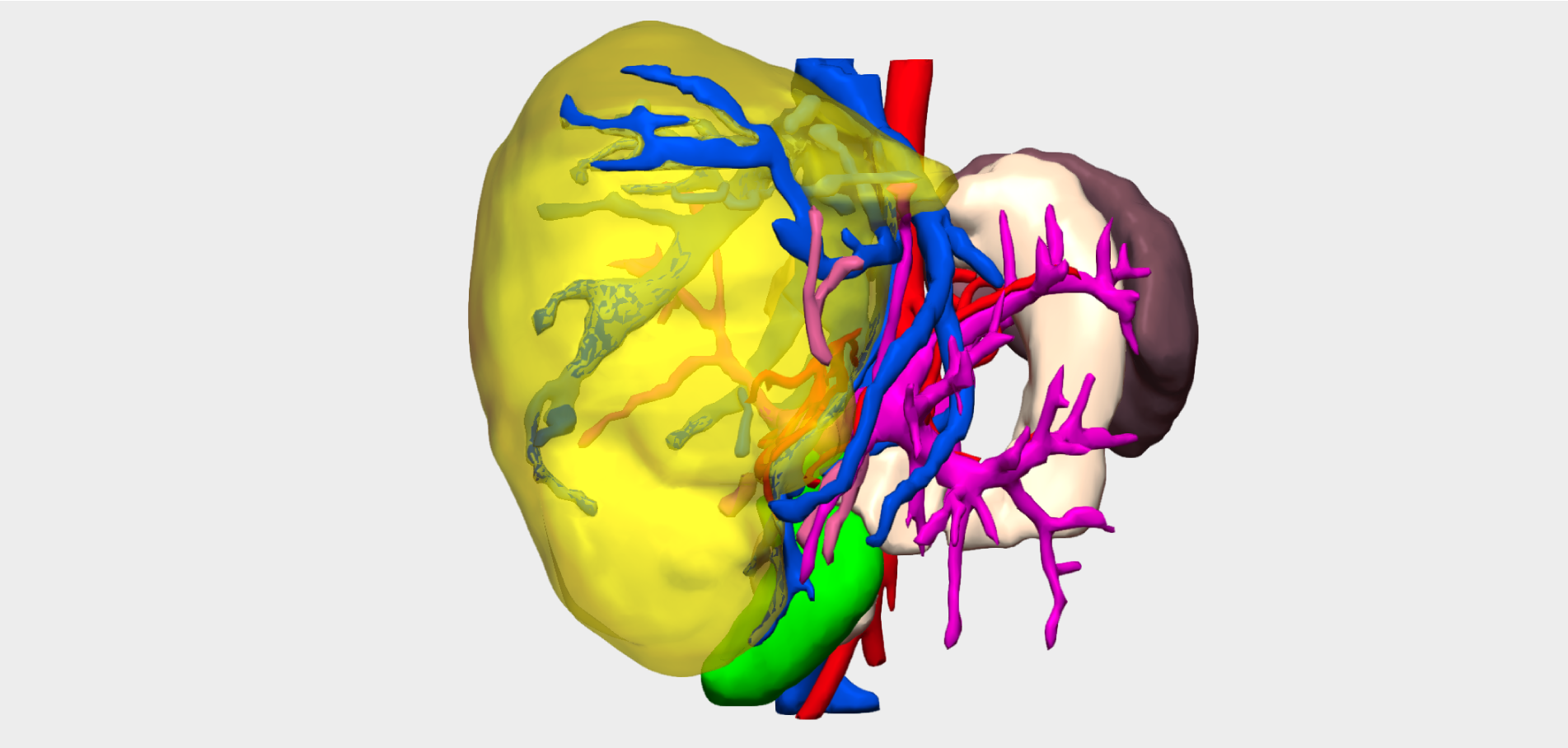
Anatomical relationships
Shows possible infiltrations/contacts between tumours and other elements.

Splenoportal axis study
Analysis of mean calibres, anatomical variants, presence of thrombosis, cavernomatosis or portosystemic shunts.

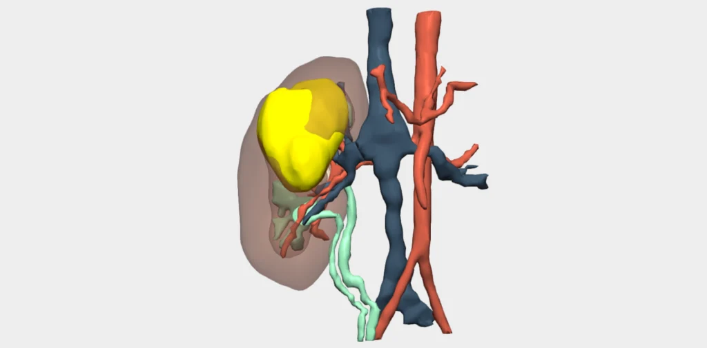
Clamping simulation
Visualise the clamping area to plan selective ischaemia.

Vascular variants
Detailed analysis of the patient’s vascular variants.
And more…
Clinical indications in Paediatric Surgery with 3D models
Especially recommended for conditions and procedures in which preoperative planning can optimise outcomes and enhance surgical safety.
PAEDIATRIC ABDOMINAL SURGERY
- Hirschsprung’s disease
- Complicated appendicitis
- Hepatobiliary pathologies
- Biliary atresia
- Choledochal cysts
- Abdominal trauma
PAEDIATRIC UROLOGICAL SURGERY
- Vesicoureteral reflux
- Congenital hydronephrosis (Pyeloureteral stenosis)
- Megaureter
- Intra-abdominal cryptorchidism
PAEDIATRIC THORACIC SURGERY
Congenital lung malformations
- Pulmonary sequestration
- Cystic adenomatoid malformation (CAM)
- Congenital lobar emphysema
Infectious diseases
- Lung abscesses
Mediastinal tumours
- Lymphoma
- Teratoma
- Intrathoracic neuroblastoma
- Bronchogenic cysts
Chest wall deformities
- Pectus excavatum
- Pectus carinatum
PAEDIATRIC ONCOLOGICAL SURGERY
Renal tumours
- Nephroblastoma (Wilms’ tumour)
- Rare tumours such as clear cell carcinoma of the kidney or rhabdoid tumours.
Soft tissue sarcomas
- Rhabdomyosarcoma and other tumours in complex locations (pelvis, thorax, retroperitoneum)
- Neuroblastoma
- Hepatoblastoma / Paediatric hepatocellular carcinoma
- Soft tissue sarcomas
Testimonials from surgeons who already use our 3D models
”Prenatal 3D reconstruction has made it possible to see the lesions directly, study their anatomical relationships, and manipulate and plan the surgery perfectly according to the size of the newborn.
Dr. Bernardo NúñezPaediatric surgeon. Parc Taulí Hospital, Sabadell, Barcelona
”Neuroblastoma surgery guided by 3D technology has already become a tangible, real advance that helps us understand the potential risks and explain them to the family and the rest of the surgical team.
Dr. Óscar GirónHead of the Oncological and Thoracic Surgery Department. Virgen de la Arrixaca University Hospital


Clinical cases in Paediatric Cancer Surgery
Learn about real success stories
Request them additionally
3D Printing models
They act as a haptic extension in the operating theatre. Each one undergoes a rigorous quality control and radiological validation process to ensure its accuracy.
- Real scale
- Haptic interpretation of anatomy
- Structure differentiation by colours
- Advanced modelling
- Sterilisable using standard methods (H₂O₂)
- Ready for rapid delivery (<72 hours)
- Intended for research and teaching purposes only

© 2026 Cella Medical Solutions