
3D models for Kidney Surgery
Urological Surgery
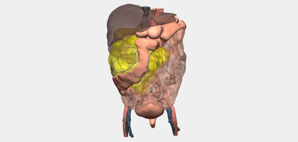
Oncological surgery for kidney cancer is the recommended treatment to remove tumours and, in some cases, the kidney itself. Cella’s 3D surgical planner provides dedicated tools to safely plan tumour resections, clamping procedures, or nephrectomies.
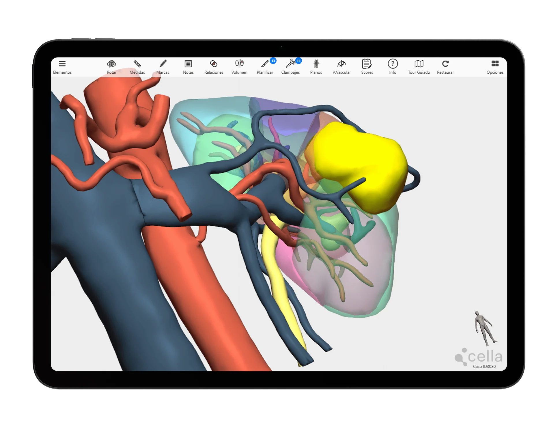
An advanced solution for planning complex kidney surgeries, enhancing clinical outcomes and enabling new surgical possibilities.
- Better understanding of the patient’s anatomy and less cognitive effort required by the surgeon to analyse structures
- Useful tool in the education and training of residents
- Less intraoperative blood loss
- Facilitates interdisciplinary communication between doctors
- Reduction in intraoperative complications
Specific tools for 3D planning in Kidney Surgery
We collaborate with urological surgeons to develop dedicated tools
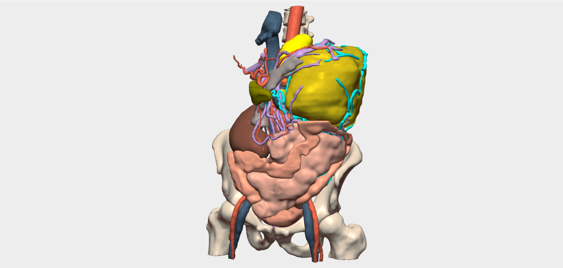
Our 3D models offer features tailored to real clinical needs, enabling personalised surgical planning for across specialties and pathologies.
Clamping simulation
Visualise the clamping area to plan selective ischaemia.
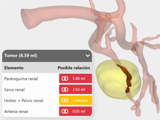
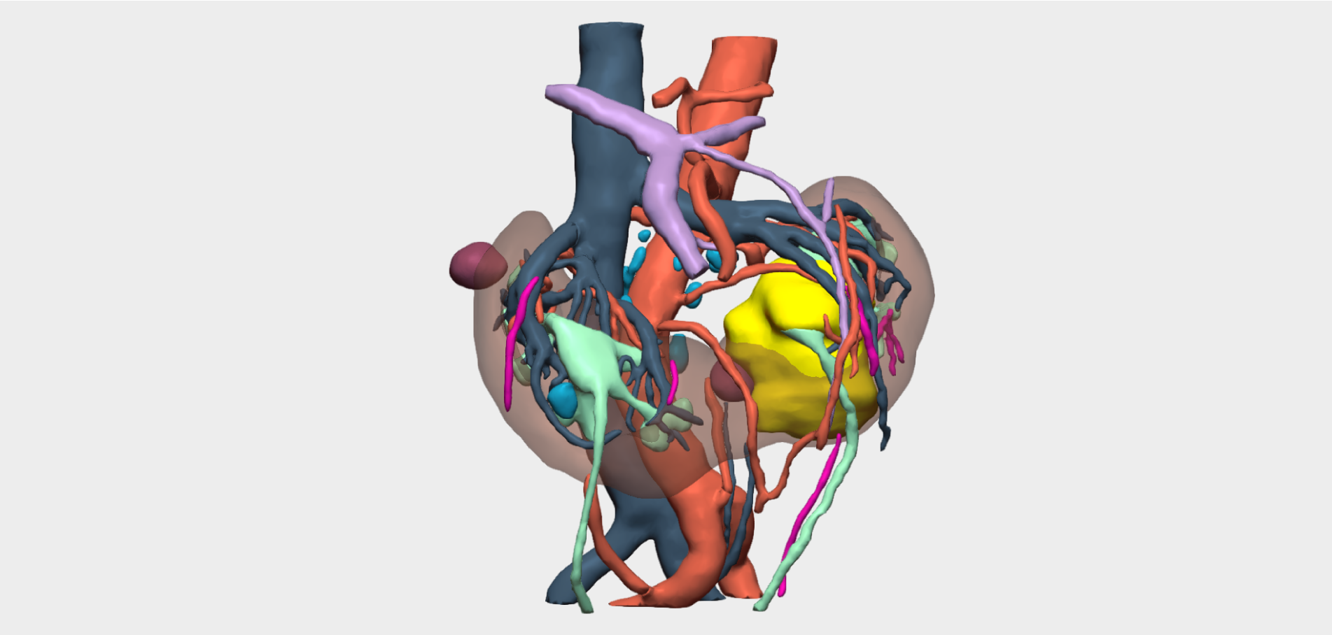
Relationship analysis
Shows possible infiltrations/contacts between the tumour and other elements.
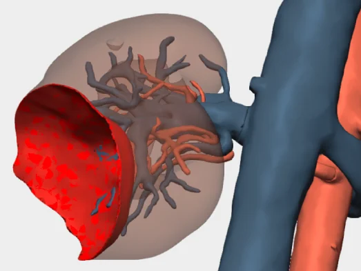
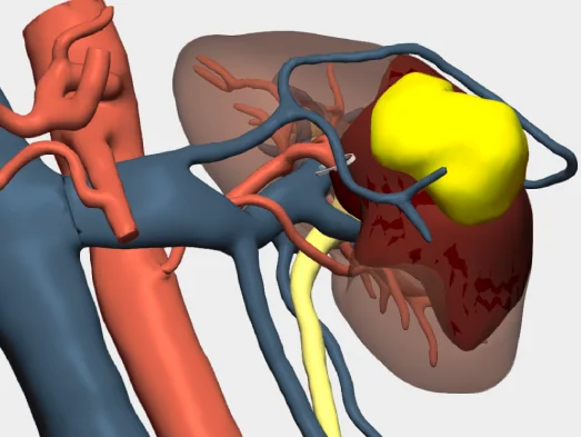
Visualisation of the surgical bed
Shows a simulation of the surgical bed after lesion removal.
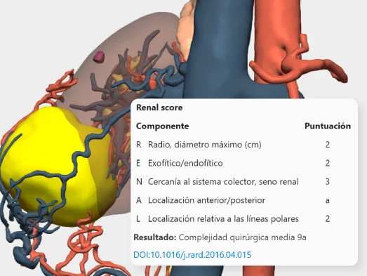
Scores
Displays the Renal Score and Padua Score in detail using a table of contents.
And more…
Clinical indications in Kidney Surgery with 3D models
Especially recommended for conditions and procedures in which preoperative planning can optimise outcomes and enhance surgical safety.
Kidney tumours
- Renal cell carcinoma (RCC)
- Renal angiomyolipoma
- Oncocytoma
- Cystic tumours
Renal cystic pathologies
- Simple renal cyst
- Polycystic kidney disease (autosomal dominant and autosomal recessive)
- Pseudocysts
Inflammatory and infectious diseases
- Acute pyelonephritis
- Chronic pyelonephritis
- Renal or perinephric abscess
- Renal tuberculosis
Obstructive pathology
- Hydronephrosis
- Renal lithiasis
- Pyeloureteral stenosis
Vascular pathologies
- Renal artery stenosis
- Renal artery aneurysm
- Renal vein thrombosis
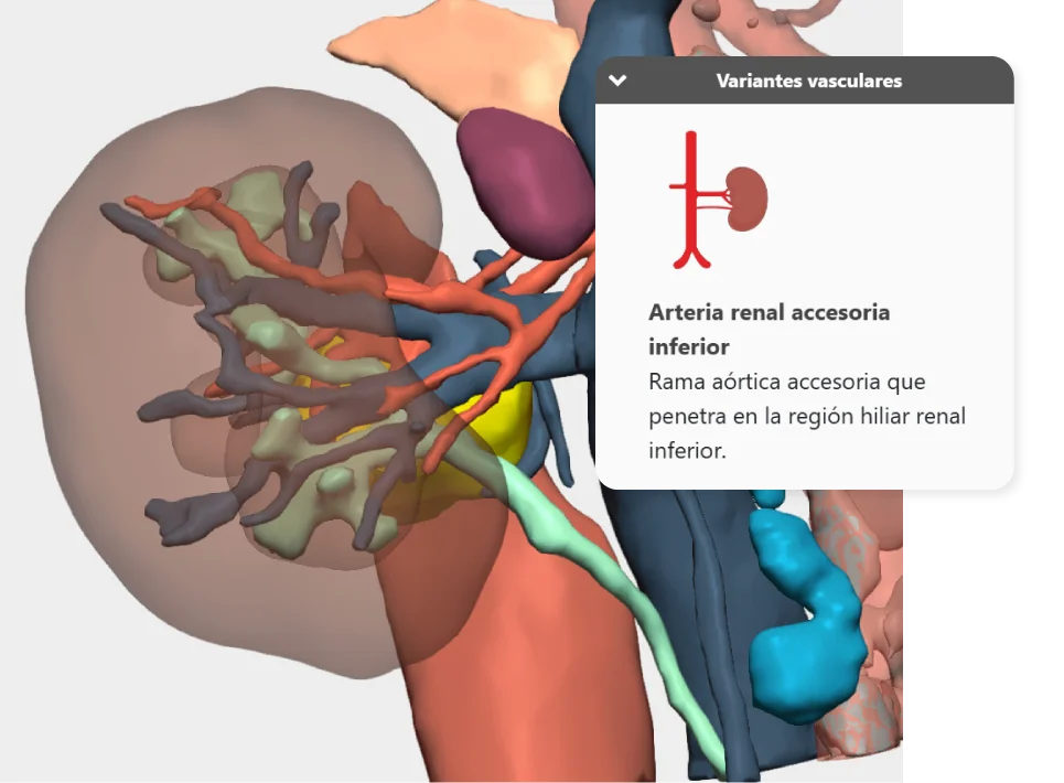
- Vascular variants
Congenital disorders
- Horseshoe kidney
- Congenital polycystic kidney disease
- Pyelocaliceal duplication
Testimonials from surgeons who already use our 3D models
”The 3D model in robotic surgery is a tool, I would dare to say essential, because it assists in vascular and urinary navigation of the renal tumour and allows for the customisation of surgical treatment.
Dr. Vital HeviaMedical Director and Head of the Kidney and Retroperitoneum Unit. ROC Clinic / HM Hospitals, Madrid
”3D virtual models provide us with easier visual information and detailed knowledge of anatomy. They also have advanced tools for assessing tumour margins, relationships with vital structures, and vascular territories.
Dr. Ignacio CastillónHead of the Urology Unit, Nuestra Señora del Rosario Hospital, Madrid


Clinical cases in Kidney Cancer Surgery
Learn about real success stories
Request them additionally
3D Printing models
They act as a haptic extension in the operating theatre. Each one undergoes a rigorous quality control and radiological validation process to ensure its accuracy.
- Real scale
- Haptic interpretation of anatomy
- Structure differentiation by colours
- Advanced modelling
- Sterilisable using standard methods (H₂O₂)
- Ready for rapid delivery (<72 hours)
- Intended for research and teaching purposes only

© 2026 Cella Medical Solutions