
3D models for Head and Neck Surgery
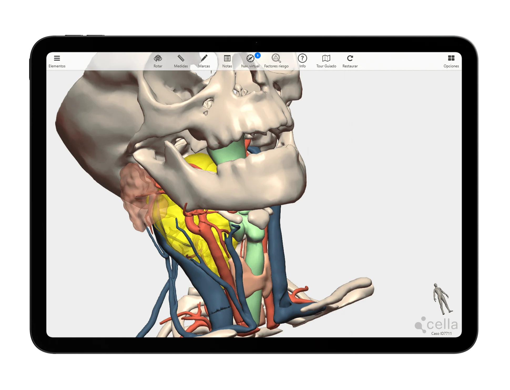
Cella Surgical Planner
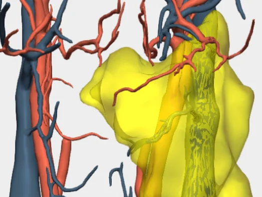
Head and neck tumours are characterised by their impact on structures such as the larynx, pharynx and salivary glands, and their proximity to nerves, vessels and vital functions like swallowing and phonation. 3D models for head and neck surgery help to delineate tumour margins, preserving nerves such as the facial nerve and facilitate reconstruction of the affected area.
An advanced solution for planning complex head and neck surgeries, enhancing clinical outcomes and enabling new surgical possibilities.
- Helps accurately identify critical structures such as nerves and blood vessels
- Improves communication among the medical team, helping to define the optimal surgical procedure
- Allows the feasibility and complexity of the surgery to be assessed
- Improves communication with the patient, facilitating their understanding of their condition and the surgical procedure
Specific tools for 3D planning of Head and Neck Surgeries
We collaborate with head and neck surgeons to develop dedicated tools
Our 3D models offer features tailored to real clinical needs, enabling personalised surgical planning for across specialties and pathologies.
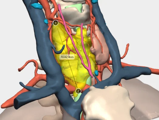
Measure distances
Measure the distance between points of interest or the diameter of elements such as vascularisation.
Surgical margins
Simulate the 5-10-15 mm safety margin for surgical resection of the tumour.
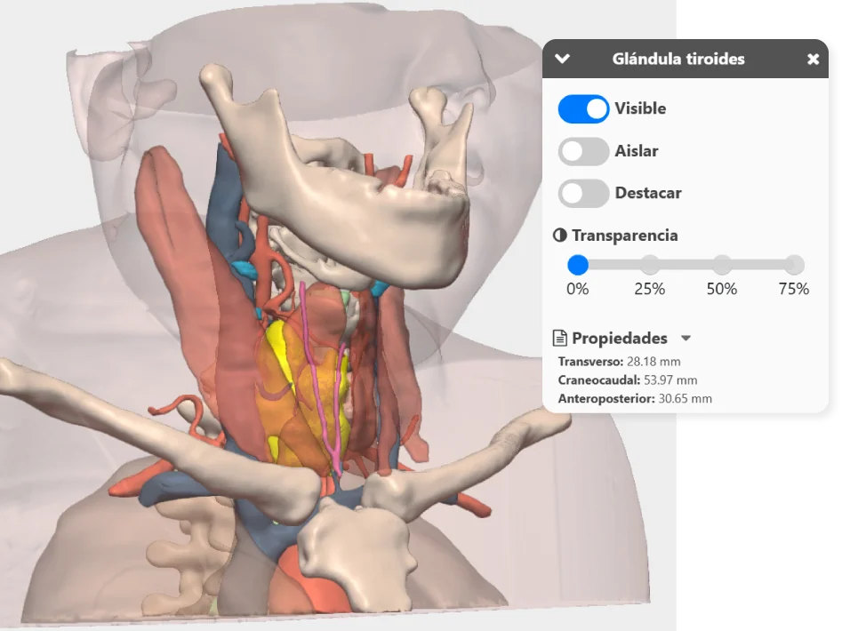
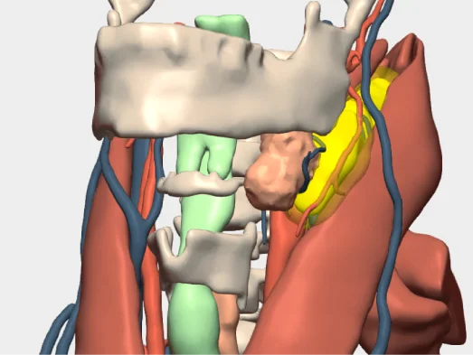
Anatomical relationships
Study the relationship of the tumour to the vascularisation, trachea, thyroid, oesophagus, and other structures.
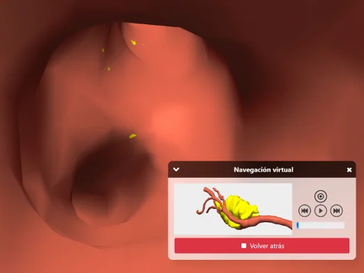
Intraductal and intravascular navigation
Visualise the interior of vascularisation or other elements to analyse tumour invasion.
And more…
Clinical indications in Head and Neck Surgery with 3D models
Especially recommended for conditions and procedures in which preoperative planning can optimise outcomes and enhance surgical safety.
Salivary gland pathology
- Salivary gland tumours
- Parotid gland
- Submandibular and sublingual glands
- Minor salivary gland tumours
- Sialolithiasis (salivary stones)
- Chronic sialadenitis
Thyroid and parathyroid pathology
- Goitre and thyroid nodules
- Thyroid cancer
- Hyperparathyroidism
Tumours and conditions of the oral cavity, oropharynx and larynx
Oral cavity
- Malignant tumours (squamous cell carcinoma of the tongue, gums, oral mucosa) and benign tumours (papillomas, fibromas).
- Tongue disorders: macroglossia, lingual cysts, congenital lesions.
Oropharynx
- Oropharyngeal carcinomas (tonsils, base of tongue, soft palate) associated or not with HPV.
- Peritonsillar or retropharyngeal abscesses in children and adults.
Larynx
- Laryngeal carcinoma (glottic, supraglottic, subglottic)
- Benign lesions (polyps, nodules, laryngeal papillomatosis)
Pathologies of the cervical wall and jaw
- Craniofacial trauma
- Mandibular osteonecrosis
- Resections for bone tumours
Nasal and paranasal sinus disorders
- Complicated chronic sinusitis
- Paranasal sinus tumours
- Cysts and cystic lesions
Pathologies of the skull base
Skull base tumours
- Malignant lesions
- Chordoma
- Chondrosarcoma
- Meningioma
- Benign lesions
- Ossifying fibromas
- Epidermoid cysts in craniofacial regions
Transoral or transfacial surgery
Congenital and cystic pathologies of the neck
- Branchial cysts and fistulas
- Thyroglossal cyst
- Lymphatic cyst (cystic hygroma)
Request them additionally
3D Printing models
They act as a haptic extension in the operating theatre. Each one undergoes a rigorous quality control and radiological validation process to ensure its accuracy.
- Real scale
- Haptic interpretation of anatomy
- Structure differentiation by colours
- Advanced modelling
- Sterilisable using standard methods (H₂O₂)
- Ready for rapid delivery (<72 hours)
- Intended for research and teaching purposes only

© 2026 Cella Medical Solutions