
3D models for Esophagogastric Surgery
General and Digestive System Surgery
Esophageal and Gastric Surgery presents significant challenges due to the anatomical complexity of the region and the high risk of complications, such as anastomotic leaks and injury to vital structures 3D models improve the identification of critical areas and support the planning of safer, more personalised procedures for each patient.
An advanced solution for planning complex esophageal and gastric surgeries, enhancing clinical outcomes and enabling new surgical possibilities.
Esophageal Surgery
- Improved lymph node recovery
- Greater precision in preoperative staging
- Better identification of neighbouring structures
Gastric Surgery
- Reduction in operating time and blood loss
- Improvement in preoperative staging of gastric cancer
- Support in surgical planning
- In-depth understanding of complex anatomy
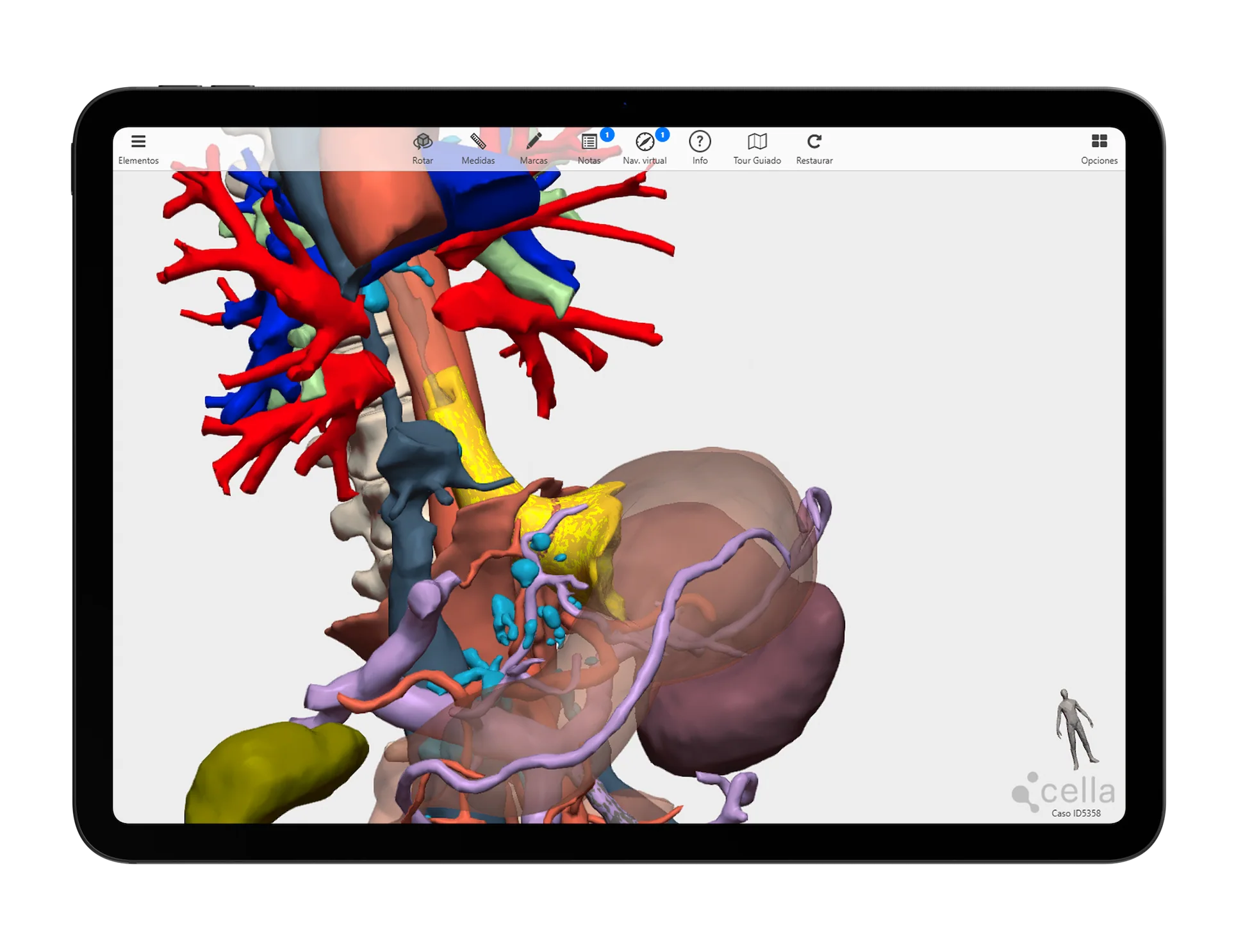
Specific tools for 3D planning in Esophagogastric Surgery
We collaborate with esophagus and gastric surgeons to develop dedicated tools
Our 3D models offer features tailored to real clinical needs, enabling personalised surgical planning for across specialties and pathologies.
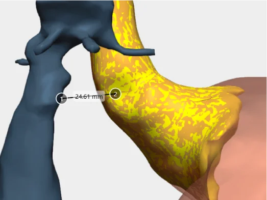
Measure distances
Measure the distance between points of interest or the diameter of elements such as tumours or vascularisation.
Surgical margins
Simulate the 5-10-15 mm safety margin for surgical resection of the tumour.
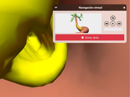
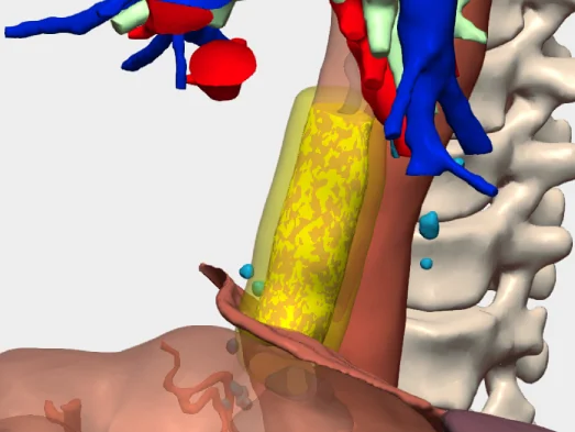
Intraductal and intravascular virtual navigation
Visualise the inside of the oesophagus, stomach or vascularisation to analyse tumour invasion.
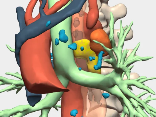
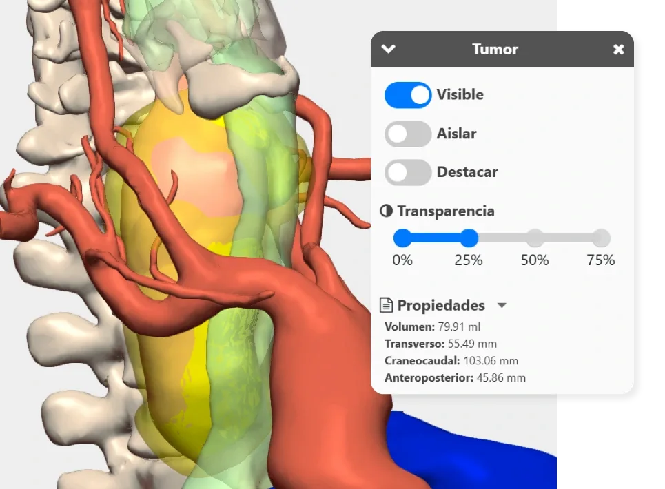
Relationship analysis
Studies how the tumour relates to adjacent elements.
And more…
Clinical indications in Esophagogastric Surgery with 3D models
Especially recommended for conditions and procedures in which preoperative planning can optimise outcomes and enhance surgical safety.
Esophagus
- Esophageal cancer
- Esophagitis
- Achalasia
- Hiatus hernia
- Esophageal diverticula
- Esophageal varices
- Rupture and tear syndromes
Stomach
- Stomach cancer (gastric adenocarcinoma)
- GIST (gastrointestinal stromal tumours)
- Gastric ulcer
- Gastritis
- Ménétrier’s disease
- Gastric polyps
- Gastric volvulus
- Gastrostomy and post-surgical complications
Testimonials from surgeons who already use our 3D models
”3D technology has been extremely helpful in countless surgical procedures, although its systematic application in the field of oesophageal and gastric surgery has yet to be discovered.
Dr. Yannko GonzálezUniversity Hospital of Torrevieja, Alicante
Clinical cases in Esophagus and Stomach Cancer Surgery
Learn about real success stories
Request them additionally
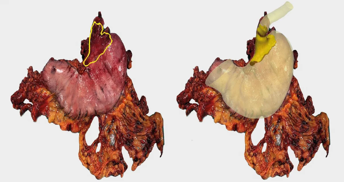
3D Printing models
They act as a haptic extension in the operating theatre. Each one undergoes a rigorous quality control and radiological validation process to ensure its accuracy.
- Real scale
- Haptic interpretation of anatomy
- Structure differentiation by colours
- Advanced modelling
- Sterilisable using standard methods (H₂O₂)
- Ready for rapid delivery (<72 hours)
- Intended for research and teaching purposes only

© 2026 Cella Medical Solutions